In a paper accepted for publication in the American Institute of Physics' journal Review of Scientific Instruments,
a team of researchers describes an advanced experimental system that
can generate attosecond bursts of extreme ultraviolet light. Such pulses
are the shortest controllable light pulses available to science. With
these pulses, according to the researchers, it's possible to measure the
dynamics of electrons in matter in real-time. Advances in attosecond
science may enable scientists to verify theories that describe how
matter behaves at a fundamental level, how certain important chemical
reactions – such as photosynthesis – work. Additional advances may
eventually lead to the control of chemical reactions.
"Understanding how matter works at the level of its electrons is
likely to lead to new scientific tools and to novel technologies," said
Felix Frank, of Imperial College in London and one of the authors on the
paper. "In the future, this knowledge could help us to make better
drugs, more efficient solar cells, and other things we can't yet
foresee."
The researchers were able to produce these pulses by a process called
high harmonic generation (HHG). The fundamental technology driving
their setup is a high-power femtosecond laser system (femtoseconds are
three orders of magnitude longer than attoseconds). The near infrared
femtosecond laser pulses are corralled through a waveguide and a series
of specialized mirrors, causing them to be compressed in time. With
their waveforms precisely controlled, these compressed pulses are then
focused into a gas target, creating an attosecond burst of extreme
ultraviolet radiation. The experimental system developed by the
researchers is able to accurately measure the attosecond
pulses and deliver them to a variety of experiments in conjugation with
other precisely synchronized laser pulses. "Though it incorporates many
novel features, our system builds on a decade of research conducted by
physics groups around the world," said John Tisch, lead scientist
developing the technology at Imperial College.
Saturday, June 30, 2012


Tuesday, June 12, 2012
Flashes of light out of the mirror
(Phys.org) -- A team of the Laboratory of Attosecond physics at
the Max Planck Institute of Quantum Optics developed an alternative way
of generating attosecond flashes of light.
Electrons at a glass surface send out flashes of light with durations of only a few attoseconds when they come under the influence of high-intensity laser pulses. One attosecond is one part in a billion of one part in a billion of a second. In the electric field of the laser, the electrons at the surface start to oscillate. Hereby the ultrashort attosecond flashes of light are generated. The team at the Laboratory of Attosecond Physics (LAP) at the Max Planck Institute of Quantum Optics (MPQ) in Garching has now advanced this innovative method. It has the potential to replace the current procedure of the generation of attosecond flashes of light. Presently these flashes are generated by electrons in noble gases. But the scientists are sure, that their method of the generation of attosecond flashes of light at surfaces has some advantages (Physical Review Letters).
Flashes of light with attosecond duration enable observations in a world yet widely unknown – the microcosm. With their help the first images of the extremely fast motion of electrons became possible. The short bursts of light are usually generated by the use of noble gas atoms. The electrons of these atoms absorb the energy of the laser light and subsequently emit it again in the form of ultrashort flashes of light. It holds: The shorter the burst of light, the sharper the images out of the microcosm.
But there are other ways of generating these short bursts of light. A team at the Laboratory of Attosecond Physics (LAP) at the Max Planck Institute of Quantum Optics (MPQ) in Garching has now advanced one of these methods. The scientists shot a laser pulse with a duration of only 8 femtoseconds and a power of 16 terawatts onto a glass target, which thereby turned into a relativistically oscillating mirror. One femtosecond corresponds to one part in a million of one part in a billion of one second and 16 terrawatt correspond to the power of round about 1000 nuclear power stations.
The 8 femtosecond laser pulse consisted of only 3 optical cycles and hence 3 cycles of its electric field. As soon as this electric field hits the glass surface a relativistic plasma forms. This means, that the electrons at the surface are accelerated out of the solid to velocities close to the speed of light and subsequently are decelerated and sent back to the surface again, as soon as the electric field changes its polarization. Thereby the electrons form an oscillating mirror. During the reflection at this moving mirror the pulsed laser light is converted from the near infrared spectral region down to the extreme ultraviolet (XUV, down to a wavelength of 17 nanometer) part of the spectrum. Hereby even shorter flashes of light with a duration in the attosecond regime are generated. These flashes of light occur as isolated bursts or trains of pulses, if filtered appropriately. Comparison with simulations of the method show that the ultrashort flashes of light have durations of around 100 attoseconds.
Compared to the conventional method of attosecond pulse generation these new flashes of light possess a higher number of photons and are hence more intense than their predecessors. This higher intensity allows for the splitting of these isolated bursts into two parts which enables the observation of processes in the microcosm with two attosecond flashes of light. This in turn permits a higher resolution than achievable up to now with the use of an attosecond burst in combination with a longer femtosecond laser pulse.
For ultrashort imaging this means that images with a greater richness of detail will become achievable in the future.
More information: P. Heissler, et al. Few-cycle driven relativistically oscillating plasma mirrors - a source of intense, isolated attosecond pulses, Phys. Rev. Lett. 108, 235003 (2012)
Electrons at a glass surface send out flashes of light with durations of only a few attoseconds when they come under the influence of high-intensity laser pulses. One attosecond is one part in a billion of one part in a billion of a second. In the electric field of the laser, the electrons at the surface start to oscillate. Hereby the ultrashort attosecond flashes of light are generated. The team at the Laboratory of Attosecond Physics (LAP) at the Max Planck Institute of Quantum Optics (MPQ) in Garching has now advanced this innovative method. It has the potential to replace the current procedure of the generation of attosecond flashes of light. Presently these flashes are generated by electrons in noble gases. But the scientists are sure, that their method of the generation of attosecond flashes of light at surfaces has some advantages (Physical Review Letters).
Flashes of light with attosecond duration enable observations in a world yet widely unknown – the microcosm. With their help the first images of the extremely fast motion of electrons became possible. The short bursts of light are usually generated by the use of noble gas atoms. The electrons of these atoms absorb the energy of the laser light and subsequently emit it again in the form of ultrashort flashes of light. It holds: The shorter the burst of light, the sharper the images out of the microcosm.
But there are other ways of generating these short bursts of light. A team at the Laboratory of Attosecond Physics (LAP) at the Max Planck Institute of Quantum Optics (MPQ) in Garching has now advanced one of these methods. The scientists shot a laser pulse with a duration of only 8 femtoseconds and a power of 16 terawatts onto a glass target, which thereby turned into a relativistically oscillating mirror. One femtosecond corresponds to one part in a million of one part in a billion of one second and 16 terrawatt correspond to the power of round about 1000 nuclear power stations.
The 8 femtosecond laser pulse consisted of only 3 optical cycles and hence 3 cycles of its electric field. As soon as this electric field hits the glass surface a relativistic plasma forms. This means, that the electrons at the surface are accelerated out of the solid to velocities close to the speed of light and subsequently are decelerated and sent back to the surface again, as soon as the electric field changes its polarization. Thereby the electrons form an oscillating mirror. During the reflection at this moving mirror the pulsed laser light is converted from the near infrared spectral region down to the extreme ultraviolet (XUV, down to a wavelength of 17 nanometer) part of the spectrum. Hereby even shorter flashes of light with a duration in the attosecond regime are generated. These flashes of light occur as isolated bursts or trains of pulses, if filtered appropriately. Comparison with simulations of the method show that the ultrashort flashes of light have durations of around 100 attoseconds.
Compared to the conventional method of attosecond pulse generation these new flashes of light possess a higher number of photons and are hence more intense than their predecessors. This higher intensity allows for the splitting of these isolated bursts into two parts which enables the observation of processes in the microcosm with two attosecond flashes of light. This in turn permits a higher resolution than achievable up to now with the use of an attosecond burst in combination with a longer femtosecond laser pulse.
For ultrashort imaging this means that images with a greater richness of detail will become achievable in the future.
More information: P. Heissler, et al. Few-cycle driven relativistically oscillating plasma mirrors - a source of intense, isolated attosecond pulses, Phys. Rev. Lett. 108, 235003 (2012)
Friday, June 08, 2012
Tabletop laser-like device creates coherent multicolor beams of ultraviolet, T- and X-rays
For the first time, researchers have produced a coherent, laser-like, directed beam of light that simultaneously streams ultraviolet light, X-rays, and all wavelengths in between.
One of the few light sources to successfully produce a coherent beam that includes X-rays, this new technology is the first to do so using a setup that fits on a laboratory table.
An international team of researchers, led by engineers from the NSF Engineering Research Center (ERC) for EUV Science and Technology, reports their findings in the June 8, 2012, issue of Science.
By focusing intense pulses of infrared light--each just a few optical cycles in duration--into a high-pressure gas cell, the researchers converted part of the original laser energy into a coherent super-continuum of light that extends well into the X-ray region of the spectrum.
The X-ray burst that emerges has much shorter wavelengths than the original laser pulse, which will make it possible to follow the tiniest, fastest physical processes in nature, including the coupled dance of electrons and ions in molecules as they undergo chemical reactions, or the flow of charges and spins in materials.
"This is the broadest spectral-bandwidth, coherent-light source ever generated," says engineering and physics professor Henry Kapteyn of JILA at the University of Colorado at Boulder, who led the study with fellow JILA professor Margaret Murnane and research scientist Tenio Popmintchev, in collaboration with researchers from the Vienna University of Technology, Cornell University and the University of Salamanca.
"It definitely opens up the possibility to probe the shortest space and time scales relevant to any process in our natural world other than nuclear or fundamental particle interactions," Kapteyn adds. The breakthrough builds upon earlier discoveries from Murnane, Kapteyn and their colleagues to generate laser-like beams of light across a broad spectrum of wavelengths.
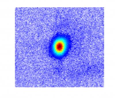 |
| This picture shows an actual image of a coherent (laser-like) X-ray beam. In contrast to the incoherent (light-bulb-like) light emitted in all directions from a Roentgen X-ray tube, the X-rays produced by high harmonic generation emerge as well-directed, laser-like, beams. |
"Just as a violin or guitar string will emit harmonics of its fundamental sound tone when plucked strongly, an atom can also emit harmonics of light when plucked violently by a laser pulse," adds Murnane. "The laser pulse first plucks electrons from the atoms, before driving them back again where they can collide with the atoms from which they came. Any excess energy is emitted as high-energy ultraviolet photons."
Like many phenomena, when HHG was first discovered, there was little science to explain it, and it was considered more a curious phenomenon than a potentially useful light source. After years of work, scientists eventually understood how very high harmonics were emitted, however there was one major challenge that most researchers gave up on--for most wavelengths in the X-ray region, the output HHG beams were extremely weak.
Murnane, Kapteyn and their students realized that there might be a chance to overcome that challenge and turn HHG into a useful X-ray light source--the tabletop-scale X-ray laser that has been a goal for laser science since shortly after the laser was first demonstrated in 1960.
"This was not an easy task," says Murnane. "Unlike a laser--which gets more intense as more energy is pumped into the system--in HHG, if the laser hits the atoms too hard, too many electrons are liberated from the gas atoms, and those electrons cause the laser light to speed up. If the speed of the laser and X-rays do not match, there is no way to combine the many X-ray waves together to create a bright output beam, since the X-ray waves from different gas atoms will interfere destructively."
Popmintchev and JILA graduate student Ming-Chang Chen worked out conditions that enable X-ray waves from many atoms in the gas to interfere constructively. The key was to use a relatively long-wavelength, mid-infrared laser and a high pressure gas cell that also guides the laser light. The resulting bright, X-ray beams maintain the coherent, directed beam qualities of the laser that drives the process.
The HHG process is effective only when the atoms are hit "hard and fast" by the laser pulses, with durations nearing 10-14 seconds--a fundamental limit representing just a few oscillations of the electromagnetic fields. Murnane and Kapteyn pioneered the technology for generating such light pulses in the 1990s, and used those lasers to develop and utilize HHG-based light sources in the extreme-ultraviolet (EUV) region of the spectrum in the 2000s. However, while researchers were using those lasers and the HHG technique to measure ever-shorter duration light pulses, they were stymied in how to make coherent light at shorter wavelengths in the more penetrating X-ray region of the spectrum.
The new paper in Science, under lead author and senior research associate Popmintchev, demonstrates that breakthrough, showing that the understanding of the HHG process the researchers developed is broadly valid.
"We would have never found this if we hadn't sat down and thought about what happens overall during HHG, when we change the wavelength of the laser driving it, what parameters have to be changed to make it work," added Kapteyn. "The amazing thing is that the physics seem to be panning out even over a very broad range of parameters. Usually in science you find a scaling rule that prevents you from making a dramatic jump, but in this case, we were able to generate 1.6 keV - each X-ray photon was generated from more than 5,000 infrared photons."
When the researchers first started to work with ultrafast, mid-infrared lasers just a few years ago, they actually made a step backwards and generated bright extreme-ultraviolet light of longer wavelengths than they used to achieve in the lab.
"However, we discovered a new regime that helped us to realize, just on paper, that we could make this giant step forward towards much shorter electromagnetic wavelengths and generate bright, laser-like, soft and hard X-rays," adds Popmintchev. "What the experiments were suggesting back then looked too good to be true! It seemed that Mother Nature has combined together, in the most simple and beautiful way, all the microscopic and macroscopic physics. Now, we are already at X-ray wavelengths as short as roughly 7.7 angstroms, and we do not know the limit."
To truly control the beam of photons, the researchers needed to understand the HHG process at the atomic level and how X-rays emitted from individual atoms combine to form a coherent beam of light.
That understanding combines microscopic and macroscopic models of the HHG process with the fact that those interactions occur at very high intensity in a dynamically changing medium. The development of such a conceptual understanding took the last decade to develop.
The result was the realization that there is no fundamental limit to the energy of the photons that can be generated using the HHG process. To obtain higher-energy photons, the system paradoxically begins with laser light using lower energy photons--specifically, mid-infrared lasers.
The JILA researchers demonstrated the validity of that principle in their labs in Colorado, but to achieve their breakthrough, the researchers traveled to Vienna with their beam-generating setup. There, they used a laser developed by co-author Andrius Baltuška and colleagues at the Vienna University of Technology--the world's most-intense ultrashort-pulse laser operating in the mid-infrared, with a wavelength of four microns.
"Thirty years ago, people were saying we could make a coherent X-ray source, but it would have to be an X-ray laser, and we'd need an atomic bomb as the energy source to pump it," said Deborah Jackson, the program officer who oversees the ERC's grant. "Now, we have these guys who understand the science fundamentals well enough to introduce new tricks for efficiently extracting energetic photons, pulling them out at X-ray wavelengths . . . and it's all done on a table-top!"
In addition to achieving the high energy, the increasingly broad spectrum opens a range of new applications.
"In an experiment using such a source, one energy region from the beam will correspond with one element, another with another element, and so on to simultaneously look at atoms across entire molecules, and that will allow us to see how charge moves from one part of a molecule to another as a chemical reaction is happening," adds Kapteyn. "It'll take us awhile to learn how to use this, but it's very exciting."
More information: "Bright Coherent Ultrahigh Harmonics in the keV X-ray Regime from Mid-Infrared Femtosecond Lasers," by T. Popmintchev et al, Science, 2012.
Wednesday, April 18, 2012
Looking through the glass transition on an ultrafast timescale
Glass is one of civilization's most valuable and versatile materials. To scientists, it's also one of the most intriguing, because it displays properties of both solids and liquids.
Glass is a non-crystalline solid that transforms into a liquid when it's heated to the so-called glass-transition temperature. When glass approaches this critical temperature—which is between 970 to 1,100 degrees Fahrenheit for the most common type of glass—it is simultaneously composed of fluid, flowing regions and solid-like, rigid domains.
For decades, scientists have been trying to understand exactly how glass behaves, at the molecular level, as it approaches the transition temperature. Now a research group led by University of Michigan chemist Kevin J. Kubarych has applied ultrafast spectroscopy to observe the fastest molecular motions of a liquid hovering just above its glass transition temperature.
"Progress in demystifying the glass transition can have a wide impact in many other fields, including predicting optical and mechanical properties of polymers and understanding crowded cellular environments of living organisms," said Kubarych, an assistant professor of chemistry.
Working with U-M chemistry graduate students John King and Matthew Ross, Kubarych found that even on the time scale of picoseconds there are signatures of "dynamic arrest": The molecules become locked into their positions and long-range motion grinds to a near halt, though, structurally, the glass is indistinguishable from a liquid.
Typically, these effects are observed on much slower time scales of seconds, minutes, or even longer. A paper summarizing the research was published online April 9 in Physical Review Letters. King is the first author of the paper.
More information: Ultrafast α-Like Relaxation of a Fragile Glass-Forming Liquid Measured Using Two-Dimensional Infrared Spectroscopy
Sunday, April 01, 2012
New technique lights up the creation of holograms
 The mechanism of full-color holography The major difference between the researchers’ technique and ordinary holography is the use of a prism to adjust the light beam’s angle of incidence in combination with a thin silver film to produce the surface plasmons. When the angle of incidence is controlled appropriately, the silver film is excited to produce surface plasmons, which in turn cause the white light to reach the hologram in the three primary colors of red, green and blue, producing a floating full-color stereoscopic image. Although only still images can be obtained at present, Kawata is planning to improve the current system to enable movie imaging based on the same principle in the future.
Thursday, March 29, 2012
Ultrafast laser pulses shed light on elusive superconducting mechanism
An international team that includes University of British Columbia physicists has used ultra-fast laser pulses to identify the microscopic interactions that drive high-temperature superconductivity.
In the experiment, to be outlined this Friday in the journal Science, electrons in a prototypical copper-oxide superconductor were excited by extremely short 100-femtosecond laser pulses.
As the material's electrons relax back to an equilibrium state, they release their excess energy via deformation of the superconductor's atomic lattice (phonons) or perturbation of its magnetic correlations (spin fluctuations).
The researchers were able to capture very fine grained data on the speed of the relaxation process and its influence on the properties of the superconducting system, showing that the high-critical temperature of these compounds can be accounted for by purely electronic (magnetic) processes.
"This new technique offers us our best window yet on the interactions that govern the formation of these elusive superconducting properties--both across time and across a wide range of characteristic energies," says UBC Associate Professor Andrea Damascelli, Canada Research Chair in Electronic Structure of Solids with the Department of Physics and Astronomy and the UBC Quantum Matter Institute.
"We're now able to begin to disentangle the different interactions that contribute to this fascinating behavior."
Superconductivity--the phenomenon of conducting electricity with no resistance--occurs in some materials at very low temperatures. High-temperature cuprate superconductors are capable of conducting electricity without resistance at temperatures as high as -140 degrees Celsius.
The key mechanism that allows the carriers to flow without resistance in superconductors stems from an effective pairing between electrons. In conventional metallic superconductors, this pairing mechanism is well understood as phonon-mediated. In copper-oxides, the nature of the low-resistance interaction between the electrons has remained a mystery.
"This breakthrough in the understanding of the puzzling properties of copper-oxides paves the way to finally solving the mystery of high-temperature superconductivity and revealing the key knobs for engineering new superconducting materials with even higher transition temperatures," says the paper's lead author Claudio Giannetti, a researcher with Italy's Università Cattolica del Sacro Cuore and visiting professor at UBC's Quantum Matter Institute.
The international collaboration also involved contributions from Japanese, Swiss and American researchers.
More information: "Disentangling the Electronic and Phononic Glue in a High-Tc Superconductor," by S. Dal Conte, et al., Science.
In the experiment, to be outlined this Friday in the journal Science, electrons in a prototypical copper-oxide superconductor were excited by extremely short 100-femtosecond laser pulses.
As the material's electrons relax back to an equilibrium state, they release their excess energy via deformation of the superconductor's atomic lattice (phonons) or perturbation of its magnetic correlations (spin fluctuations).
The researchers were able to capture very fine grained data on the speed of the relaxation process and its influence on the properties of the superconducting system, showing that the high-critical temperature of these compounds can be accounted for by purely electronic (magnetic) processes.
"This new technique offers us our best window yet on the interactions that govern the formation of these elusive superconducting properties--both across time and across a wide range of characteristic energies," says UBC Associate Professor Andrea Damascelli, Canada Research Chair in Electronic Structure of Solids with the Department of Physics and Astronomy and the UBC Quantum Matter Institute.
"We're now able to begin to disentangle the different interactions that contribute to this fascinating behavior."
Superconductivity--the phenomenon of conducting electricity with no resistance--occurs in some materials at very low temperatures. High-temperature cuprate superconductors are capable of conducting electricity without resistance at temperatures as high as -140 degrees Celsius.
The key mechanism that allows the carriers to flow without resistance in superconductors stems from an effective pairing between electrons. In conventional metallic superconductors, this pairing mechanism is well understood as phonon-mediated. In copper-oxides, the nature of the low-resistance interaction between the electrons has remained a mystery.
"This breakthrough in the understanding of the puzzling properties of copper-oxides paves the way to finally solving the mystery of high-temperature superconductivity and revealing the key knobs for engineering new superconducting materials with even higher transition temperatures," says the paper's lead author Claudio Giannetti, a researcher with Italy's Università Cattolica del Sacro Cuore and visiting professor at UBC's Quantum Matter Institute.
The international collaboration also involved contributions from Japanese, Swiss and American researchers.
More information: "Disentangling the Electronic and Phononic Glue in a High-Tc Superconductor," by S. Dal Conte, et al., Science.
Thursday, March 22, 2012
NIF facility fires record laser shot into target chamber
(PhysOrg.com) -- The National Ignition Facility (NIF) at Lawrence Livermore National Laboratory in California has set a new record for a laser shot. This past week, its combined 192 lasers fired a single 1.875-megajoule shot into an empty test chamber. After passing through the last of its focusing lens, the shot reached 2.03 megajoules, making it the first 2 megajoule ultraviolet laser.
Prior to this achievement, the most the facility had managed to coax out of the laser, the world’s largest, was 1.6 megajoules. Also, the new record shows that the NIF laser is capable of producing more than it was designed for, which was 1.8 megajoules. It also proved that it was capable of doing so without damaging its parts, allowing for another shot a day and a half later, which is important, because one of the goals for the laser is to get it to fire off shots at 15 per second eventually. That’s what researchers think will be needed to produce power economically from the laser system.
The ultimate goal of the NIF is to figure out a way to use a laser to produce nuclear fusion in a way that gets more energy out than is put in. Currently, that goal is still a ways off. Thus far, engineers at the project haven't even reached the break-even or ignition point, though they expect that to occur sometime this year. Tweaking the laser to produce more than it was designed for is a step in that direction. The NIF facility was designed to produce a fusion reaction by imploding hydrogen isotope pellets using the huge laser. To that end, the team has made steady progress. When the project first began eighteen months ago, it had just one percent of conditions in place that are believed necessary to achieve the ignition point. They have improved that mark to ten percent and it’s because the pace has picked up dramatically in recent months that they believe they will achieve the ignition point sometime over the next six months, which is when the original ignition campaign was slated to end.
Because the facility is funded by the US nuclear weapons complex, there has been debate about whether it would ever be used to prove or disprove the idea that lasers could be used to create nuclear fusion to produce electric power. Having the laser break records doesn’t really resolve that argument in the short term, but it might in the long run if it does eventually show that electricity could be created economically using such a process.
More information: The National Ignition Facility (NIF): https://lasers.llnl.gov/
Prior to this achievement, the most the facility had managed to coax out of the laser, the world’s largest, was 1.6 megajoules. Also, the new record shows that the NIF laser is capable of producing more than it was designed for, which was 1.8 megajoules. It also proved that it was capable of doing so without damaging its parts, allowing for another shot a day and a half later, which is important, because one of the goals for the laser is to get it to fire off shots at 15 per second eventually. That’s what researchers think will be needed to produce power economically from the laser system.
The ultimate goal of the NIF is to figure out a way to use a laser to produce nuclear fusion in a way that gets more energy out than is put in. Currently, that goal is still a ways off. Thus far, engineers at the project haven't even reached the break-even or ignition point, though they expect that to occur sometime this year. Tweaking the laser to produce more than it was designed for is a step in that direction. The NIF facility was designed to produce a fusion reaction by imploding hydrogen isotope pellets using the huge laser. To that end, the team has made steady progress. When the project first began eighteen months ago, it had just one percent of conditions in place that are believed necessary to achieve the ignition point. They have improved that mark to ten percent and it’s because the pace has picked up dramatically in recent months that they believe they will achieve the ignition point sometime over the next six months, which is when the original ignition campaign was slated to end.
Because the facility is funded by the US nuclear weapons complex, there has been debate about whether it would ever be used to prove or disprove the idea that lasers could be used to create nuclear fusion to produce electric power. Having the laser break records doesn’t really resolve that argument in the short term, but it might in the long run if it does eventually show that electricity could be created economically using such a process.
More information: The National Ignition Facility (NIF): https://lasers.llnl.gov/
Thursday, March 15, 2012
Scientists reveal inner workings of magnets, a finding that could lead to faster computers
Using a light source that creates X-ray pulses only one quadrillionth of a second in duration, the Boulder team was able to observe how magnetism in nickel and iron atoms works, and they found that each metal behaves differently. One quadrillionth of a second is a million times faster than one billionth of a second.
The results of the study were published online this week by the Proceedings of the National Academy of Sciences. Six of the study’s 19 co-authors are located at CU-Boulder.
Many technology experts believe that next-generation computer disk drives will use optically-assisted magnetic recording to achieve much higher drive capacities, according to NIST scientist Tom Silva, who worked with CU-Boulder physics professors Margaret Murnane and Henry Kapteyn on the research. However, many questions remain about how the delivery of optical energy to the magnetic system can be optimized for maximum drive performance. And this finding could help researchers answer some of their questions.
“The discovery that iron and nickel are fundamentally different in their interaction with light at ultrafast time scales suggests that the magnetic alloys in hard drives could be engineered to enhance the delivery of the optical energy to the spin system,” Silva said.
Magnetism exists because all of the “spins” in a magnet -- each of which is like a very small bar magnet with a north and south pole -- are lined up to point in the same direction, much like members of a marching band who are moving in unison, explained Murnane, who also is a fellow of JILA, a joint institute of CU-Boulder and NIST.
“The powerful laser pulse scrambles the magnetic spins in the metal, as if the members of the marching band started moving in different directions across the football field, causing the magnetization to rapidly disappear within a mere fifty quadrillionths of a second, a process known as ultrafast demagnetization,” Murnane said.
While ultrafast demagnetization has been a well-known phenomenon since its discovery in 1996, the CU and NIST researchers saw for the first time that different kinds of spins in metal scramble on different time scales. Until now, it was assumed that all the spins in a metal alloy behaved in the same way due to a powerful quantum mechanical effect known as the exchange interaction, which lines up all the individual spins in the same direction.
“What we have seen for the first time is that the iron spins and the nickel spins react to light in different ways, with the iron spins being mixed up by light much more readily than the nickel spins,” said Silva. “In the end, the exchange interaction still pulls the two spin systems back into synchronization after a few quadrillionths of a second. Seeing such a difference was only possible by taking advantage of the extremely fast X-ray technology developed at the University of Colorado and elsewhere.”
The laser technology used in the experiment, known as “high harmonic generation,” can generate laser-like beams of X-rays that span a wide portion of the electromagnetic spectrum, including the spectral region where nickel and iron interact very strongly with X-rays.
The results of the study were published online this week by the Proceedings of the National Academy of Sciences. Six of the study’s 19 co-authors are located at CU-Boulder.
Many technology experts believe that next-generation computer disk drives will use optically-assisted magnetic recording to achieve much higher drive capacities, according to NIST scientist Tom Silva, who worked with CU-Boulder physics professors Margaret Murnane and Henry Kapteyn on the research. However, many questions remain about how the delivery of optical energy to the magnetic system can be optimized for maximum drive performance. And this finding could help researchers answer some of their questions.
“The discovery that iron and nickel are fundamentally different in their interaction with light at ultrafast time scales suggests that the magnetic alloys in hard drives could be engineered to enhance the delivery of the optical energy to the spin system,” Silva said.
Magnetism exists because all of the “spins” in a magnet -- each of which is like a very small bar magnet with a north and south pole -- are lined up to point in the same direction, much like members of a marching band who are moving in unison, explained Murnane, who also is a fellow of JILA, a joint institute of CU-Boulder and NIST.
“The powerful laser pulse scrambles the magnetic spins in the metal, as if the members of the marching band started moving in different directions across the football field, causing the magnetization to rapidly disappear within a mere fifty quadrillionths of a second, a process known as ultrafast demagnetization,” Murnane said.
While ultrafast demagnetization has been a well-known phenomenon since its discovery in 1996, the CU and NIST researchers saw for the first time that different kinds of spins in metal scramble on different time scales. Until now, it was assumed that all the spins in a metal alloy behaved in the same way due to a powerful quantum mechanical effect known as the exchange interaction, which lines up all the individual spins in the same direction.
“What we have seen for the first time is that the iron spins and the nickel spins react to light in different ways, with the iron spins being mixed up by light much more readily than the nickel spins,” said Silva. “In the end, the exchange interaction still pulls the two spin systems back into synchronization after a few quadrillionths of a second. Seeing such a difference was only possible by taking advantage of the extremely fast X-ray technology developed at the University of Colorado and elsewhere.”
The laser technology used in the experiment, known as “high harmonic generation,” can generate laser-like beams of X-rays that span a wide portion of the electromagnetic spectrum, including the spectral region where nickel and iron interact very strongly with X-rays.
Saturday, March 10, 2012
Making sharper X-rays
 A photograph of the custom-built laser chamber, which receives simultaneously a pulse of helium gas and a pulse from RIKEN’s free-electron laser, which is known as the SPring-8 Compact SASE Source (SCSS). The resulting blue-green superfluorescence is visible.
A variety of imaging technologies rely on light with short wavelengths because it allows very small structures to be resolved. However, light sources which produce short, extreme ultraviolet or x-ray wavelengths often have unstable emission wavelength and timing. Now, by illuminating a gas with a powerful laser, a research team in Japan has demonstrated a light source that may solve many of these problems. The research was published by Mitsuru Nagasono, from the RIKEN SPring-8 Center in Harima, and his colleagues from RIKEN and three other institutes.
More information: Nagasono, M., et al. Observation of free-electron-laser-induced collective spontaneous emission (superfluorescence). Physical Review Letters 107, 193603 (2011).
Friday, March 09, 2012
Femtolaser Pulse Creates 3-D Nanostructures
A new fabrication process using femtosecond lasers creates 3-D nanostructures in materials, an essential step toward creating invisibility cloaks and other advanced materials that bend light in unusual ways.
Researchers in Eric Mazur's laboratory at the Harvard School of Engineering and Science (SEAS) fired a femtosecond laser, which releases incredibly bright flashes of light that last 5 x 10^-14, s, at a glass slide coated with a mixture of silver nitrate, water and PVP, a water-soluble polymer. The laser blast changes the electrical, physical and optical properties of the slide, and photoreduces the silver ions on the slide into nanocrystals of silver metal suspended on the polymer.
Previous attempts to create a 3-D structure failed because the coating was not quite right. When the researchers used only the silver nitrate and water, there was no lattice support for the silver atoms, they said.
"Normally, when people use femtosecond lasers in fabrication, they’re creating a woodpile structure: something stacked on something else, being supported by something else. If you want to make an array of silver dots, however, they can’t float in space," Mazur said. Ethanol and the PVP polymer were added to the solution to provide support to the structure, but reactions were fast and uncontrollable. Removing the ethanol solved the problem entirely.
"What was most surprising about it was how simple it is. It was a matter of using less," Mazur said.
The new fabrication process advances nanoscale metal lithography into three dimensions, and does it at a resolution high enough to be practical for metamaterials.
"This work demonstrates that we can create silver dots that are disconnected in X, Y, and Z," said Kevin Vora, a graduate student working on the project. "There’s no other technique that feasibly allows you to do that."
The work, which was supported by the Air Force Office of Scientific Research, is described in Applied Physics Letters.
Researchers in Eric Mazur's laboratory at the Harvard School of Engineering and Science (SEAS) fired a femtosecond laser, which releases incredibly bright flashes of light that last 5 x 10^-14, s, at a glass slide coated with a mixture of silver nitrate, water and PVP, a water-soluble polymer. The laser blast changes the electrical, physical and optical properties of the slide, and photoreduces the silver ions on the slide into nanocrystals of silver metal suspended on the polymer.
Previous attempts to create a 3-D structure failed because the coating was not quite right. When the researchers used only the silver nitrate and water, there was no lattice support for the silver atoms, they said.
"Normally, when people use femtosecond lasers in fabrication, they’re creating a woodpile structure: something stacked on something else, being supported by something else. If you want to make an array of silver dots, however, they can’t float in space," Mazur said. Ethanol and the PVP polymer were added to the solution to provide support to the structure, but reactions were fast and uncontrollable. Removing the ethanol solved the problem entirely.
"What was most surprising about it was how simple it is. It was a matter of using less," Mazur said.
The new fabrication process advances nanoscale metal lithography into three dimensions, and does it at a resolution high enough to be practical for metamaterials.
"This work demonstrates that we can create silver dots that are disconnected in X, Y, and Z," said Kevin Vora, a graduate student working on the project. "There’s no other technique that feasibly allows you to do that."
The work, which was supported by the Air Force Office of Scientific Research, is described in Applied Physics Letters.
Thursday, March 08, 2012
First-ever images of atoms moving in a molecule captured
 Researchers at Ohio State University and Kansas State University have captured the first-ever images of atoms moving in a molecule. Shown here is molecular nitrogen. The researchers used an ultrafast laser to knock one electron from the molecule, and recorded the diffraction pattern that was created when the electron scattered off the molecule. The image highlights any changes the molecule went through during the time between laser pulses: one quadrillionth of a second. The constituent atoms' movement is shown as a measure of increasing angular momentum, on a scale from dark blue to pink, with pink showing the region of greatest momentum. Credit: Image courtesy of Cosmin Blaga, Ohio State University.
Key to the experiment, which appears in this week's issue of the journal Nature, is the researchers' use of the energy of a molecule's own electron as a kind of "flash bulb" to illuminate the molecular motion.
The team used ultrafast laser pulses to knock one electron out of its natural orbit in a molecule. The electron then fell back toward the molecule scattered off of it, analogous to the way a flash of light scatters around an object, or a water ripple scatters in a pond.
Principal investigator Louis DiMauro of Ohio State University said that the feat marks a first step toward not only observing chemical reactions, but also controlling them on an atomic scale.
"Through these experiments, we realized that we can control the quantum trajectory of the electron when it comes back to the molecule, by adjusting the laser that launches it," said DiMauro, who is a professor of physics at Ohio State. "The next step will be to see if we can steer the electron in just the right way to actually control a chemical reaction."
A standard technique for imaging a still object involves shooting the object with an electron beam – bombarding it with millions of electrons per second. The researchers' new single-electron quantum approach allowed them to image rapid molecular motion, based on theoretical developments by the paper's coauthors at Kansas State University.
A technique called laser induced electron diffraction (LIED) is commonly used in surface science to study solid materials. Here, the researchers used it to study the movement of atoms in a single molecule.
The molecules they chose to study were simple ones: nitrogen, or N2, and oxygen, or O2. N2 and O2 are common atmospheric gases, and scientists already know every detail of their structure, so these two very basic molecules made a good test case for the LIED method.
In each case, the researchers hit the molecule with laser light pulses of 50 femtoseconds, or quadrillionths of a second. They were able to knock a single electron out of the outer shell of the molecule and detect the scattered signal of the electron as it re-collided with the molecule.
DiMauro and Ohio State postdoctoral researcher Cosmin Blaga likened the scattered electron signal to the diffraction pattern that light forms when it passes through slits. Given only the diffraction pattern, scientists can reconstruct the size and shape of the slits. In this case, given the diffraction pattern of the electron, the physicists reconstructed the size and shape of the molecule – that is, the locations of the constituent atoms' nuclei.
The key, explained Blaga, is that during the brief span of time between when the electron is knocked out of the molecule and when it re-collides, the atoms in the molecules have moved. The LIED method can capture this movement, "similar to making a movie of the quantum world," he added.
Beyond its potential for controlling chemical reactions, the technique offers a new tool to study the structure and dynamics of matter, he said. "Ultimately, we want to really understand how chemical reactions take place. So, long-term, there would be applications in materials science and even chemical manufacturing."
"You could use this to study individual atoms," DiMauro added, "but the greater impact to science will come when we can study reactions between more complex molecules. Looking at two atoms – that's a long way from studying a more interesting molecule like a protein."
Wednesday, February 08, 2012
With single laser pulses on single molecules
 Nowadays, large laser systems provide ultra-short light pulses of very high intensity which – in principle – allow the imaging of matter and its dynamics on atomic scales, down to a single molecule or a virus. However, current methods fall short in efficiency to overlap a target molecule in a deterministic way. Physicists around Prof. Tobias Schätz have now found a possible way out. Using the well proven concept of ion traps they store a single molecule at a precisely known position and then hit it in a deterministic way with single laser pulses that are provided by the Laboratory for Attosecond Physics at MPQ (Nature Physics). Though still restricted to pulses in the UV range this method makes it possible to resolve the internal dynamics of a single molecular ion consisting of a magnesium ion and a hydrogen atom. “However, this scheme could become a standard technique for investigating large biomolecules, if X-ray laser pulses can be applied”, Tobias Schätz points out.
At present there is no satisfying method for investigating the structure of large and complex molecules, e.g., proteins. The standard technique is based on the diffraction of X-rays in crystals and fails in this case, because many biological molecules are difficult or impossible to crystallize. Diffraction experiments on single molecules with low-intensity sources require long exposure times in order to reach the number of about 1013 photons which is necessary to achieve an image. This leads to radiation damage of the target particle and, furthermore, excludes the temporal resolution required to analyze short-lived intermediate products or fast structural changes.
A new generation of X-ray femtosecond lasers promises to overcome these limitations. Light pulses comprising a huge number of photons within a period of a few femtoseconds produce images of a single molecule before the radiation damage becomes visible (1 femtosecond corresponds to 10^-15 seconds). In addition, the beam diametre of the laser has to be focused down to the size of a molecule, about a tenth of a micrometre. This has been accomplished already. The challenge is now to prepare a single molecule so reliable that it can be deterministically placed within the laser pulse.
In the past couple of decades ion traps have provided unique control capabilities for charged particles. An ion trap is basically a small vacuum chamber containing four electrodes which are switched rapidly between minus and plus, at frequencies in the radio frequency range (107 Hertz). Under the influence of these quickly changing electrical fields a single ion (i.e. an electrically charged atom), which has been cooled down to very low temperatures, gets trapped in the centre of the chamber. Isolated from the environment the 'floating' ion can remain there for hours. If several ions are guided into the trap a structured pattern evolves, due to their mutual repulsion. This reminds of a solid state crystal, yet, the lattice sites are much easier to resolve, since the distances between the ions are a 100 000 times larger.
In contrast to atomic ions molecules are much more difficult to trap because they cannot directly be cooled. The MPQ team has now resumed to a trick: they embed the molecule into a crystal formed by cooled atomic ions. The experimental set up consists of two ion traps connected in series. In the first trap the molecular ion is prepared in a photochemical reaction from magnesium and hydrogen, i.e., each molecule consists of a positively charged magnesium ion and a hydrogen atom. These molecular ions are transferred into a second ion trap already filled with atomic magnesium ions which have arranged themselves into a regular pattern, keeping a distance of 10 micrometres from each other. In this very cold environment also the single molecule comes to a rest and replaces one of the atoms in the ion crystal. Whereas the atomic ions emit light by fluorescence, the lattice site occupied with the molecule remains dark. The absolute position of the molecular ion can then be deduced by detecting the fluorescence light of its neighbors with an accuracy of less than a micrometer.
Now the conditions are set for hitting the single molecule with a femtosecond laser pulse at a probability of almost 100 percent. In the beginning the molecule finds itself in a vibrational ground state. With a first so-called pump pulse it gets excited into a state in which its two components – the magnesium ion and the hydrogen atom – oscillate with a period of 30 femtoseconds. A short time later a second pulse ‘probes’ in which phase of the oscillation cycle the molecule is at that very moment. At the turning point of the oscillation, after 15 femtoseconds, the distance between the particles has reached its maximum. If the probe pulse hits the molecule at that time, the dissociation probability is particularly high. The breaking of the chemical bond is signaled by the disappearance of a non fluorescing dark spot.
“In our experiment we should be able to provide the molecules at the rate of the laser pulses, i.e., about a hundred per second”, Tobias Schätz explains. “So each time a molecule is damaged by radiation it can be replaced by an identical one. As we vary the delay between pump- and probe-pulse we can resolve the vibrational dynamics of the bi-atomic molecule. This is due to the fact that the laser pulse duration of a few femtoseconds is much shorter than the molecular oscillation cycle.”
The experiment described here is a demonstration of the feasibility and the potential of the new technique which for the first time combines ion traps with classical pump-probe set-ups. The use of X-rays instead of UV-pulses will make it possible to apply the technique to biomolecules which in nature often show up as charged articles. The high intensity and the short duration of the X-Ray pulses will allow obtaining useful information on the structure of the molecule before it suffers from radiation damage. In the future experiments of that kind could be the key to investigate single complex molecules with the necessary precision and efficiency.
More information: Steffen Kahra, et al., Controlled delivery of single molecules into ultra-short laser pulses: a molecular conveyor belt, Nature Physics, AOP, 5 February 2012.
Thursday, January 26, 2012
First atomic X-ray laser created
 Scientists working at the U.S. Department of Energy's (DOE) SLAC National Accelerator Laboratory have created the shortest, purest X-ray laser pulses ever achieved, fulfilling a 45-year-old prediction and opening the door to a new range of scientific discovery.
This artist's conception illustrates how the new atomic hard X-ray laser is created. A powerful X-ray laser pulse from SLAC National Accelerator Laboratory's Linac Coherent Light Source comes up from the lower-left corner (shown as green) and hits a neon atom (center). This intense incoming light energizes an electron from an inner orbit (or shell) closest to the neon nucleus (center, brown), knocking it totally out of the atom (upper-left, foreground). In some cases, an outer electron will drop down into the vacated inner orbit (orange starburst near the nucleus) and release a short-wavelength, high-energy (i.e., "hard") X-ray photon of a specific wavelength (energy/color) (shown as yellow light heading out from the atom to the upper right along with the larger, green LCLS light). X-rays made in this manner then stimulate other energized neon atoms to do the same, creating a chain-reaction avalanche of pure X-ray laser light amplified by a factor of 200 million. While the LCLS X-ray pulses are brighter and more powerful, the neon atomic hard X-ray laser pulses have one-eighth the duration and a much purer light color. This new laser will enable more precise investigations into ultrafast processes and chemical reactions than had been possible before, ultimately opening the door to new medicines, devices and materials. Credit: Illustration by Gregory M. Stewart, SLAC National Accelerator Laboratory
World's most powerful X-ray laser creates two-million-degree matter
The experiments were carried out at SLAC's Linac Coherent Light Source (LCLS), whose rapid-fire laser pulses are a billion times brighter than those of any X-ray source before it. Scientists used those pulses to flash-heat a tiny piece of aluminum foil, creating what is known as "hot dense matter," and took the temperature of this solid plasma—about 2 million degrees Celsius. The whole process took less than a trillionth of a second.
"The LCLS X-ray laser is a truly remarkable machine," said Sam Vinko, a postdoctoral researcher at Oxford University and the paper's lead author. "Making extremely hot, dense matter is important scientifically if we are ultimately to understand the conditions that exist inside stars and at the center of giant planets within our own solar system and beyond."
Scientists have long been able to create plasma from gases and study it with conventional lasers, said co-author Bob Nagler of SLAC, an LCLS instrument scientist. But no tools were available for doing the same at solid densities that cannot be penetrated by conventional laser beams.
"The LCLS, with its ultra-short wavelengths of X-ray laser light, is the first that can penetrate a dense solid and create a uniform patch of plasma—in this case a cube one-thousandth of a centimeter on a side—and probe it at the same time," Nagler said.
The resulting measurements, he said, will feed back into theories and computer simulations of how hot, dense matter behaves. This could help scientists analyze and recreate the nuclear fusion process that powers the sun.
"Those 60 hours when we first aimed the LCLS at a solid were the most exciting 60 hours of my entire scientific career," said Justin Wark, leader of the Oxford group. "LCLS is really going to revolutionize the field, in my view."
"The LCLS X-ray laser is a truly remarkable machine," said Sam Vinko, a postdoctoral researcher at Oxford University and the paper's lead author. "Making extremely hot, dense matter is important scientifically if we are ultimately to understand the conditions that exist inside stars and at the center of giant planets within our own solar system and beyond."
Scientists have long been able to create plasma from gases and study it with conventional lasers, said co-author Bob Nagler of SLAC, an LCLS instrument scientist. But no tools were available for doing the same at solid densities that cannot be penetrated by conventional laser beams.
"The LCLS, with its ultra-short wavelengths of X-ray laser light, is the first that can penetrate a dense solid and create a uniform patch of plasma—in this case a cube one-thousandth of a centimeter on a side—and probe it at the same time," Nagler said.
The resulting measurements, he said, will feed back into theories and computer simulations of how hot, dense matter behaves. This could help scientists analyze and recreate the nuclear fusion process that powers the sun.
"Those 60 hours when we first aimed the LCLS at a solid were the most exciting 60 hours of my entire scientific career," said Justin Wark, leader of the Oxford group. "LCLS is really going to revolutionize the field, in my view."
Sunday, January 22, 2012
Physicists cool semiconductor by laser light
Researchers at the Niels Bohr Institute have combined two worlds – quantum physics and nano physics, and this has led to the discovery of a new method for laser cooling semiconductor membranes. Semiconductors are vital components in solar cells, LEDs and many other electronics, and the efficient cooling of components is important for future quantum computers and ultrasensitive sensors. The new cooling method works quite paradoxically by heating the material. Using lasers, researchers cooled membrane fluctuations to minus 269 degrees C. The results are published in the scientific journal, Nature Physics.
 The experiments themselves are carried out in this vacuum chamber. When the laser light hits the membrane, some of the light is reflected and some is absorbed and leads to a small heating of the membrane. The reflected light is reflected back again via a mirror in the experiment so that the light flies back and forth in this space and forms optical resonator (cavity). Changing the distance between the membrane and the mirror leads to a complex and fascinating interplay between the movement of the membrane, the properties of the semiconductor and the optical resonances and you can control the system so as to cool the temperature of the membrane fluctuations.
From gas to solid
Laser cooling of atoms has been practiced for several years in experiments in the quantum optical laboratories of the Quantop research group at the Niels Bohr Institute. Here researchers have cooled gas clouds of cesium atoms down to near absolute zero, minus 273 degrees C, using focused lasers and have created entanglement between two atomic systems. The atomic spin becomes entangled and the two gas clouds have a kind of link, which is due to quantum mechanics. Using quantum optical techniques, they have measured the quantum fluctuations of the atomic spin.
"For some time we have wanted to examine how far you can extend the limits of quantum mechanics – does it also apply to macroscopic materials? It would mean entirely new possibilities for what is called optomechanics, which is the interaction between optical radiation, i.e. light, and a mechanical motion," explains Professor Eugene Polzik, head of the Center of Excellence Quantop at the Niels Bohr Institute at the University of Copenhagen.
But they had to find the right material to work with.
In 2009, Peter Lodahl (who is today a professor and head of the Quantum Photonic research group at the Niels Bohr Institute) gave a lecture at the Niels Bohr Institute, where he showed a special photonic crystal membrane that was made of the semiconducting material gallium arsenide (GaAs). Eugene Polzik immediately thought that this nanomembrane had many advantageous electronic and optical properties and he suggested to Peter Lodahl's group that they use this kind of membrane for experiments with optomechanics. But this required quite specific dimensions and after a year of trying they managed to make a suitable one.
"We managed to produce a nanomembrane that is only 160 nanometers thick and with an area of more than 1 square millimetre. The size is enormous, which no one thought it was possible to produce," explains Assistant Professor Søren Stobbe, who also works at the Niels Bohr Institute.
Basis for new research
Now a foundation had been created for being able to reconcile quantum mechanics with macroscopic materials to explore the optomechanical effects.
Koji Usami explains that in the experiment they shine the laser light onto the nanomembrane in a vacuum chamber. When the laser light hits the semiconductor membrane, some of the light is reflected and the light is reflected back again via a mirror in the experiment so that the light flies back and forth in this space and forms an optical resonator. Some of the light is absorbed by the membrane and releases free electrons. The electrons decay and thereby heat the membrane and this gives a thermal expansion. In this way the distance between the membrane and the mirror is constantly changed in the form of a fluctuation.
"Changing the distance between the membrane and the mirror leads to a complex and fascinating interplay between the movement of the membrane, the properties of the semiconductor and the optical resonances and you can control the system so as to cool the temperature of the membrane fluctuations. This is a new optomechanical mechanism, which is central to the new discovery. The paradox is that even though the membrane as a whole is getting a little bit warmer, the membrane is cooled at a certain oscillation and the cooling can be controlled with laser light. So it is cooling by warming! We managed to cool the membrane fluctuations to minus 269 degrees C", Koji Usami explains.
"The potential of optomechanics could, for example, pave the way for cooling components in quantum computers. Efficient cooling of mechanical fluctuations of semiconducting nanomembranes by means of light could also lead to the development of new sensors for electric current and mechanical forces. Such cooling in some cases could replace expensive cryogenic cooling, which is used today and could result in extremely sensitive sensors that are only limited by quantum fluctuations," says Professor Eugene Polzik.
 The experiments themselves are carried out in this vacuum chamber. When the laser light hits the membrane, some of the light is reflected and some is absorbed and leads to a small heating of the membrane. The reflected light is reflected back again via a mirror in the experiment so that the light flies back and forth in this space and forms optical resonator (cavity). Changing the distance between the membrane and the mirror leads to a complex and fascinating interplay between the movement of the membrane, the properties of the semiconductor and the optical resonances and you can control the system so as to cool the temperature of the membrane fluctuations.
From gas to solid
Laser cooling of atoms has been practiced for several years in experiments in the quantum optical laboratories of the Quantop research group at the Niels Bohr Institute. Here researchers have cooled gas clouds of cesium atoms down to near absolute zero, minus 273 degrees C, using focused lasers and have created entanglement between two atomic systems. The atomic spin becomes entangled and the two gas clouds have a kind of link, which is due to quantum mechanics. Using quantum optical techniques, they have measured the quantum fluctuations of the atomic spin.
"For some time we have wanted to examine how far you can extend the limits of quantum mechanics – does it also apply to macroscopic materials? It would mean entirely new possibilities for what is called optomechanics, which is the interaction between optical radiation, i.e. light, and a mechanical motion," explains Professor Eugene Polzik, head of the Center of Excellence Quantop at the Niels Bohr Institute at the University of Copenhagen.
But they had to find the right material to work with.
In 2009, Peter Lodahl (who is today a professor and head of the Quantum Photonic research group at the Niels Bohr Institute) gave a lecture at the Niels Bohr Institute, where he showed a special photonic crystal membrane that was made of the semiconducting material gallium arsenide (GaAs). Eugene Polzik immediately thought that this nanomembrane had many advantageous electronic and optical properties and he suggested to Peter Lodahl's group that they use this kind of membrane for experiments with optomechanics. But this required quite specific dimensions and after a year of trying they managed to make a suitable one.
"We managed to produce a nanomembrane that is only 160 nanometers thick and with an area of more than 1 square millimetre. The size is enormous, which no one thought it was possible to produce," explains Assistant Professor Søren Stobbe, who also works at the Niels Bohr Institute.
Basis for new research
Now a foundation had been created for being able to reconcile quantum mechanics with macroscopic materials to explore the optomechanical effects.
Koji Usami explains that in the experiment they shine the laser light onto the nanomembrane in a vacuum chamber. When the laser light hits the semiconductor membrane, some of the light is reflected and the light is reflected back again via a mirror in the experiment so that the light flies back and forth in this space and forms an optical resonator. Some of the light is absorbed by the membrane and releases free electrons. The electrons decay and thereby heat the membrane and this gives a thermal expansion. In this way the distance between the membrane and the mirror is constantly changed in the form of a fluctuation.
"Changing the distance between the membrane and the mirror leads to a complex and fascinating interplay between the movement of the membrane, the properties of the semiconductor and the optical resonances and you can control the system so as to cool the temperature of the membrane fluctuations. This is a new optomechanical mechanism, which is central to the new discovery. The paradox is that even though the membrane as a whole is getting a little bit warmer, the membrane is cooled at a certain oscillation and the cooling can be controlled with laser light. So it is cooling by warming! We managed to cool the membrane fluctuations to minus 269 degrees C", Koji Usami explains.
"The potential of optomechanics could, for example, pave the way for cooling components in quantum computers. Efficient cooling of mechanical fluctuations of semiconducting nanomembranes by means of light could also lead to the development of new sensors for electric current and mechanical forces. Such cooling in some cases could replace expensive cryogenic cooling, which is used today and could result in extremely sensitive sensors that are only limited by quantum fluctuations," says Professor Eugene Polzik.
Monday, October 24, 2011
Plasmonics produces extreme UV light
 An international team of researchers has invented a simple way of creating ultrashort pulses of extreme ultraviolet (EUV) light. The system uses a new 3D metallic waveguide, or "nanofunnel", that coverts pulses of infrared light to EUV.
EUV light has a wavelength of around 5–50 nm, which is about 100–10 times shorter than that of visible light. As a result, ultrashort pulses of EUV light are ideal for studying fundamental physics phenomena – such as how electrons move in atoms, molecules and solids.
However, it is difficult to produce EUV radiation using conventional methods that rely on using amplified light pulses from an oscillator (a source of laser light) to ionize noble gas atoms. The electrons liberated during this process are accelerated in the light field and their surplus energy is freed as attosecond (10–18 s) pulses of light of different wavelengths. The shortest wavelengths of light can then be "filtered out" to produce a single EUV pulse – a complicated process.
Simpler way of making pulses
Now, researchers at the Korea Advanced Institute of Science and Technology (KAIST), the Max Planck Institute of Quantum Optics (MPQ) in Germany and Georgia State University (GSU) in the US have come up with a different – and much simpler – way of doing things.
The new technique works by converting femtosecond (10–15 s) infrared pulses into femtosecond EUV pulses. The process exploits surface-plasmon polaritons (SPPs), which are particle-like collective oscillations that occur when light interacts with a metal's conduction electrons.
The nanofunnel made by the KAIST-MPQ-GSU team was devised so that it concentrated incident infrared light pulses into a spot that is smaller than the wavelength of the incident light. The funnel is a metallic nanostructure made of silver that contains a hollow hole shaped like a tapered cone. The cone is just a few micrometres long and filled with xenon gas. The tip of the funnel is around 100 nm across.
The researchers sent infrared light pulses (at a rate of 75 MHz) into the funnel, which is designed so that it contains patches of metal that are positively charged, followed by patches that are negatively charged. This arrangement produces electromagnetic fluctuations on the inside walls of the funnel, which result in the creation of SPPs. These particles then travel towards the tip, where the conical shape of the funnel concentrates their fields.
"The field on the inside of the funnel can become a few hundred times stronger than the field of the incident infrared light," explains Mark Stockman of GSU. "This enhanced field results in the generation of EUV light in the Xe gas."
An important feature of the nanofunnel is that it can be produced at frequencies of up to about 75 MHz. Seung-Woo Kim, team leader at KAIST, where the experiments were carried out, adds: "Due to their short wavelength and potentially short pulse duration, EUV light pulses can be an important tool for exploring electron dynamics in atoms, molecules and solids. Electrons move very fast – on the attosecond timescale – and light flashes that are shorter than attoseconds long are therefore needed to image these particles. Although scientists routinely use attosecond light flashes for such studies, they have much lower frequencies. Our new nanofunnel could change all this."
The results are detailed in Nature Photonics.
Tuesday, September 20, 2011
Brightest gamma ray on Earth -- for a safer, healthier world
Physicists have discovered that ultra-short duration laser pulses can interact with ionised gas to give off beams that are so intense they can pass through 20 cm of lead and would take 1.5 m of concrete to be completely absorbed.
The ray could have several uses, such as in medical imaging, radiotherapy and radioisotope production for PET (positron emission tomography) scanning. The source could also be useful in monitoring the integrity of stored nuclear waste.
In addition, the laser pulses are short enough- lasting a quadrillionth of a second- to capture the response of a nucleus to stimuli, making the rays ideal for use in lab-based study of the nucleus.
The device used in the research is smaller and less costly than more conventional sources of gamma rays, which are a form of X-rays.
 The experiments were carried out on the Gemini laser in the Central Laser Facility at the Science and Technology Facilities Council's Rutherford Appleton Laboratory. Strathclyde was also joined in the research by University of Glasgow and Instituto Superior Técnico in Lisbon.
Professor Dino Jaroszynski of Strathclyde, who led the research, said: "This is a great breakthrough, which could make the probing of very dense matter easier and more extensive, and so allow us to monitor nuclear fusion capsules imploding.
"To prove this we have imaged very thin wires - 25 microns thick - with gamma rays and produced very clear images using a new method called phase-contrast imaging. This allows very weakly absorbing material to be clearly imaged. Matter illuminated by gamma rays only cast a very weak shadow and therefore are invisible. Phase-contrast imaging is the only way to render these transparent objects visible.
"It could also act as a powerful tool in medicine for cancer therapy and there is nothing else to match the duration of the gamma ray pulses, which is also why it is so bright.
"In nature, if you accelerate charged particles, such as electrons, they radiate. We trapped particles in a cavity of ions trailing an intense laser pulse and accelerated these to high energies. Electrons in this cavity also interact with the laser and pick up energy from it and oscillate wildly - much like a child being pushed on a swing. The large swinging motion and the high energy of the electrons allow a huge increase in the photon energy to produce gamma rays. This enabled the gamma ray photons to outshine any other earthbound source.
"The accelerator we use is a new type called a laser-plasma wakefield accelerator which uses high power lasers and ionised gas to accelerate charged particles to very high energies - thus shrinking a conventional accelerator, which is 100m long, to one which fits in the palm of your hand."
The peak brilliance of the gamma rays was measured to be greater than 1023 photons per second, per square milliradian, per square millimetre, per 0.1% bandwidth.
The research was supported by the Engineering and Physical Sciences Research Council, the Science and Technology Facilities Council, the Laserlab-Europe Consortium and the Extreme Light Infrastructure project. It is linked to SCAPA (Scottish Centre for the Application of Plasma-based Accelerators), which is based at Strathclyde and is run through the Scottish Universities Physics Alliance.
The research has been published in the journal Nature Physics.
The ray could have several uses, such as in medical imaging, radiotherapy and radioisotope production for PET (positron emission tomography) scanning. The source could also be useful in monitoring the integrity of stored nuclear waste.
In addition, the laser pulses are short enough- lasting a quadrillionth of a second- to capture the response of a nucleus to stimuli, making the rays ideal for use in lab-based study of the nucleus.
The device used in the research is smaller and less costly than more conventional sources of gamma rays, which are a form of X-rays.
 The experiments were carried out on the Gemini laser in the Central Laser Facility at the Science and Technology Facilities Council's Rutherford Appleton Laboratory. Strathclyde was also joined in the research by University of Glasgow and Instituto Superior Técnico in Lisbon.
Professor Dino Jaroszynski of Strathclyde, who led the research, said: "This is a great breakthrough, which could make the probing of very dense matter easier and more extensive, and so allow us to monitor nuclear fusion capsules imploding.
"To prove this we have imaged very thin wires - 25 microns thick - with gamma rays and produced very clear images using a new method called phase-contrast imaging. This allows very weakly absorbing material to be clearly imaged. Matter illuminated by gamma rays only cast a very weak shadow and therefore are invisible. Phase-contrast imaging is the only way to render these transparent objects visible.
"It could also act as a powerful tool in medicine for cancer therapy and there is nothing else to match the duration of the gamma ray pulses, which is also why it is so bright.
"In nature, if you accelerate charged particles, such as electrons, they radiate. We trapped particles in a cavity of ions trailing an intense laser pulse and accelerated these to high energies. Electrons in this cavity also interact with the laser and pick up energy from it and oscillate wildly - much like a child being pushed on a swing. The large swinging motion and the high energy of the electrons allow a huge increase in the photon energy to produce gamma rays. This enabled the gamma ray photons to outshine any other earthbound source.
"The accelerator we use is a new type called a laser-plasma wakefield accelerator which uses high power lasers and ionised gas to accelerate charged particles to very high energies - thus shrinking a conventional accelerator, which is 100m long, to one which fits in the palm of your hand."
The peak brilliance of the gamma rays was measured to be greater than 1023 photons per second, per square milliradian, per square millimetre, per 0.1% bandwidth.
The research was supported by the Engineering and Physical Sciences Research Council, the Science and Technology Facilities Council, the Laserlab-Europe Consortium and the Extreme Light Infrastructure project. It is linked to SCAPA (Scottish Centre for the Application of Plasma-based Accelerators), which is based at Strathclyde and is run through the Scottish Universities Physics Alliance.
The research has been published in the journal Nature Physics.
Saturday, September 10, 2011
Researchers at NIF moving closer to fusion ignition point
(PhysOrg.com) -- Researchers at the U.S. National Ignition Facility (NIF) report that they are growing ever closer to reaching the ignition point with their laser generated nuclear fusion project. The facility, part of the Lawrence Livermore National Laboratory has been doing research to find out if very high powered lasers could be used to create nuclear fusion that could then be used to drive steam turbines to make electricity. In related news, officials for UK companies AWE and the Rutherford Appleton Laboratory have announced that they are joining forces with the research team working on the NIF project, adding years of expertise in both nuclear fusion and laser technology.
To achieve inertial confinement fusion, researchers at the NIF project shoot multiple (192) very high powered lasers at a single pellet comprised of the hydrogen isotope deuterium, which causes it to compress to a fraction of its original size and fuse into helium atoms -releasing neutrons. The neutrons could then, in theory, be used to heat water to drive steam turbines. The only problem is, thus far, the power consumed by the lasers (some shots use more power than the whole rest of the United States) exceeds the power produced by firing them at the pellet. But, that appears to be changing.
At a meeting this past week, sponsored by London’s Royal Society, representatives from the US facility and its two new British partners met to announce the terms of agreement between them all. NIF Director Ed Moses told the group (according to the BBC) that one shot of the NIF recently produced, for just the tiniest fraction of a second, more power than all the rest of the world was consuming. And while that is certainly impressive, it’s still just a fraction of what is needed to achieve ignition; the point where a self-sustaining chain reaction occurs (required for energy gain). Moses added that he believes the group will achieve ignition within the next couple of years. Part of the reason for his optimism is the advances that have been made in high power laser diodes over the ten years since the NIF was first designed.
One possible issue the group will certainly need to discuss is the enormous number of hydrogen pellets that would be needed to feed a facility that was actually engaged in producing electricity; some estimates range to 10 pellets a second, or a million every day.
If the team is successful in its endeavor, the enormous amounts of tax dollars spent will be more than made up for in energy production. Just 1300 pounds of water, for example, could provide as much electricity as 2 million metric tons of coal.
To achieve inertial confinement fusion, researchers at the NIF project shoot multiple (192) very high powered lasers at a single pellet comprised of the hydrogen isotope deuterium, which causes it to compress to a fraction of its original size and fuse into helium atoms -releasing neutrons. The neutrons could then, in theory, be used to heat water to drive steam turbines. The only problem is, thus far, the power consumed by the lasers (some shots use more power than the whole rest of the United States) exceeds the power produced by firing them at the pellet. But, that appears to be changing.
At a meeting this past week, sponsored by London’s Royal Society, representatives from the US facility and its two new British partners met to announce the terms of agreement between them all. NIF Director Ed Moses told the group (according to the BBC) that one shot of the NIF recently produced, for just the tiniest fraction of a second, more power than all the rest of the world was consuming. And while that is certainly impressive, it’s still just a fraction of what is needed to achieve ignition; the point where a self-sustaining chain reaction occurs (required for energy gain). Moses added that he believes the group will achieve ignition within the next couple of years. Part of the reason for his optimism is the advances that have been made in high power laser diodes over the ten years since the NIF was first designed.
One possible issue the group will certainly need to discuss is the enormous number of hydrogen pellets that would be needed to feed a facility that was actually engaged in producing electricity; some estimates range to 10 pellets a second, or a million every day.
If the team is successful in its endeavor, the enormous amounts of tax dollars spent will be more than made up for in energy production. Just 1300 pounds of water, for example, could provide as much electricity as 2 million metric tons of coal.
White laser pulses with precisely tailored waveform enable control of electrons in microcosm
(PhysOrg.com) -- An expedition through the fast-paced microscopic world of atoms reveals electrons that spin around at enormous speeds and have gigantic forces are acting on them. Monitoring the ultrafast motion of these electrons requires ultrashort flashes of light. However, in order to control them, the structure of these light flashes, or light pulses, needs to be tamed as well.
This type of control over light pulses has now, for the first time, been achieved by a team of physicists lead by Eleftherios Goulielmakis and Ferenc Krausz of the Laboratory of Attosecond Physics at the Max Planck Institute of Quantum Optics (MPQ) and the Ludwig-Maximilians-University Munich in Garching, along with collaborators from the Center of Free-Electron Laser Science (DESY Hamburg) and the King Saud University (Saudi Arabia). Additionally, the researchers were able to make their pulses shorter than a complete light oscillation, thereby creating for the first time isolated sub-optical-cycle flashes of light. These novel tools allow for the precise control of electron motion in atoms and molecules.
The motion of electrons in the microcosm occurs on an attosecond time scale, where one attosecond is a billionth of a billionth of a second. On such a short scale, only light itself is able to keep up with the motion. Because of the fast oscillations of its electromagnetic field, light can act somewhat like a pair of tweezers on electrons, influencing their motions and interactions. The time it takes light, generated by modern laser sources, to complete one full oscillation amounts to around 2.6 femtoseconds, where one femtosecond is a thousand attoseconds, or one millionth of a billionth of a second.
This is the reason why light is a promising tool for controlling electron dynamics in the microcosm. Yet, before this can become reality, the light’s field oscillations have to be tamed, i.e. its field has to be precisely and completely controllable on a time scale which is shorter than one full oscillation cycle. In order to achieve this lofty goal, researchers first have to learn how to develop and perfect these extraordinary tweezers.
The international team at MPQ around Eleftherios Goulielmakis and Ferenc Krausz has now mastered a big step towards this ambitious aim, managing to sculpt wave forms of laser pulses with sub-cycle precision. Furthermore, the researchers were able to make their pulses shorter than a complete light oscillation, thereby creating for the first time isolated sub-optical-cycle flashes of light.
Extremely short and intense pulses of light with a new waveform
In order to control light pulses on a sub-cycle time scale, it is necessary to use white laser light, as it contains wavelengths (light colours) ranging from the near-ultraviolet through to the visible and all the way to the near infrared region of the electromagnetic spectrum. The physicists have created these light pulses and sent them into a newly developed “light field synthesizer”. The light field synthesizer is analogous to a sound synthesizer, as used by electronic musicians. Just as the sound synthesizer, which superimposes sound waves of different frequencies to create different sounds and beats, the light field synthesizer superimposes optical waves of different colours and phases to create various field shapes. The apparatus first splits the incident white laser light into red, yellow and blue colour channels. After manipulating the properties of the individual colours, they are recombined to form the synthesized wave form. Several components of this novel device, e.g. its mirrors and its elaborate beam splitters, were developed in the service centre of the Munich Centre for Advanced Photonics (MAP) located at the LMU.
Utilizing this technology, the scientists achieved the generation of completely new isolated waveforms. Furthermore, in doing so they managed to compose the shortest pulses ever measured in the visible spectral range, lasting only 2.1 femtoseconds. These pulses are more intense than the ones commonly afforded by current femtosecond light sources because all the energy of the electromagnetic field is confined into a tiny temporal window. Thus, also the strength of the electromagnetic forces increases.
It is precisely these powerful and specially tailored electromagnetic forces which are necessary to control electrons in atoms and molecules, as they are similar in strength to the forces occurring in such microscopic systems. However, to steer electron motion on a microscopic scale, strength is not the only prerequisite because precision is also needed. This level of desired precision is provided by the well-controlled wave forms of the synthesized light pulses.
Electronics which can be controlled with light waves comes close
Thanks to these latest results, the scientists have accomplished a major step towards the control of the microcosm. “These newly developed tools allow us to initiate, control and therefore further understand inner-atomic processes. With these devices, we can master the fine structuring of ultrashort light fields and reliably measure the newly formed light”, explains Dr. Adrian Wirth, Postdoctoral Fellow in the research team of Eleftherios Goulielmakis, leader of the ERC-research group “Attoelectronics”.
As a matter of fact, the physicists have already applied this novel technique in an experiment. By shining the newly designed light pulses onto krypton atoms, the outermost electron was ripped away within less than 700 attoseconds, marking the fastest electronic process which has been initiated by optically visible light. Similar processes can certainly be achieved in more complex systems such as molecules, solids and nano-particles.
This new technology may very well lead the way towards light-based electronics in the future. Light fields are expected to drive electrons not only in isolated systems such as atoms or molecules, but even on microscopic circuits so as to perform logic operations at unprecedented speeds” said Goulielmakis, whose group is exploring the principles of electronics on these extreme time scales. “We are progressively increasing our understanding of the principles in the microcosm and learning how to control it”, adds Ferenc Krausz.
More information: Adrian Wirth, Mohammed Th. Hassan, Ivanka Grguraš, Justin Gagnon, Antoine Moulet, Tran T. Luu, Stefan Pabst, Robin Santra, Zeyad A. Alahmed, Abdallah M. Azzeer, Vladislav S. Yakovlev, Volodymyr Pervak, Ferenc Krausz & Eleftherios Goulielmakis, Synthesized Light Transients
Science Express September 6, 2011
This type of control over light pulses has now, for the first time, been achieved by a team of physicists lead by Eleftherios Goulielmakis and Ferenc Krausz of the Laboratory of Attosecond Physics at the Max Planck Institute of Quantum Optics (MPQ) and the Ludwig-Maximilians-University Munich in Garching, along with collaborators from the Center of Free-Electron Laser Science (DESY Hamburg) and the King Saud University (Saudi Arabia). Additionally, the researchers were able to make their pulses shorter than a complete light oscillation, thereby creating for the first time isolated sub-optical-cycle flashes of light. These novel tools allow for the precise control of electron motion in atoms and molecules.
The motion of electrons in the microcosm occurs on an attosecond time scale, where one attosecond is a billionth of a billionth of a second. On such a short scale, only light itself is able to keep up with the motion. Because of the fast oscillations of its electromagnetic field, light can act somewhat like a pair of tweezers on electrons, influencing their motions and interactions. The time it takes light, generated by modern laser sources, to complete one full oscillation amounts to around 2.6 femtoseconds, where one femtosecond is a thousand attoseconds, or one millionth of a billionth of a second.
This is the reason why light is a promising tool for controlling electron dynamics in the microcosm. Yet, before this can become reality, the light’s field oscillations have to be tamed, i.e. its field has to be precisely and completely controllable on a time scale which is shorter than one full oscillation cycle. In order to achieve this lofty goal, researchers first have to learn how to develop and perfect these extraordinary tweezers.
The international team at MPQ around Eleftherios Goulielmakis and Ferenc Krausz has now mastered a big step towards this ambitious aim, managing to sculpt wave forms of laser pulses with sub-cycle precision. Furthermore, the researchers were able to make their pulses shorter than a complete light oscillation, thereby creating for the first time isolated sub-optical-cycle flashes of light.
Extremely short and intense pulses of light with a new waveform
In order to control light pulses on a sub-cycle time scale, it is necessary to use white laser light, as it contains wavelengths (light colours) ranging from the near-ultraviolet through to the visible and all the way to the near infrared region of the electromagnetic spectrum. The physicists have created these light pulses and sent them into a newly developed “light field synthesizer”. The light field synthesizer is analogous to a sound synthesizer, as used by electronic musicians. Just as the sound synthesizer, which superimposes sound waves of different frequencies to create different sounds and beats, the light field synthesizer superimposes optical waves of different colours and phases to create various field shapes. The apparatus first splits the incident white laser light into red, yellow and blue colour channels. After manipulating the properties of the individual colours, they are recombined to form the synthesized wave form. Several components of this novel device, e.g. its mirrors and its elaborate beam splitters, were developed in the service centre of the Munich Centre for Advanced Photonics (MAP) located at the LMU.
Utilizing this technology, the scientists achieved the generation of completely new isolated waveforms. Furthermore, in doing so they managed to compose the shortest pulses ever measured in the visible spectral range, lasting only 2.1 femtoseconds. These pulses are more intense than the ones commonly afforded by current femtosecond light sources because all the energy of the electromagnetic field is confined into a tiny temporal window. Thus, also the strength of the electromagnetic forces increases.
It is precisely these powerful and specially tailored electromagnetic forces which are necessary to control electrons in atoms and molecules, as they are similar in strength to the forces occurring in such microscopic systems. However, to steer electron motion on a microscopic scale, strength is not the only prerequisite because precision is also needed. This level of desired precision is provided by the well-controlled wave forms of the synthesized light pulses.
Electronics which can be controlled with light waves comes close
Thanks to these latest results, the scientists have accomplished a major step towards the control of the microcosm. “These newly developed tools allow us to initiate, control and therefore further understand inner-atomic processes. With these devices, we can master the fine structuring of ultrashort light fields and reliably measure the newly formed light”, explains Dr. Adrian Wirth, Postdoctoral Fellow in the research team of Eleftherios Goulielmakis, leader of the ERC-research group “Attoelectronics”.
As a matter of fact, the physicists have already applied this novel technique in an experiment. By shining the newly designed light pulses onto krypton atoms, the outermost electron was ripped away within less than 700 attoseconds, marking the fastest electronic process which has been initiated by optically visible light. Similar processes can certainly be achieved in more complex systems such as molecules, solids and nano-particles.
This new technology may very well lead the way towards light-based electronics in the future. Light fields are expected to drive electrons not only in isolated systems such as atoms or molecules, but even on microscopic circuits so as to perform logic operations at unprecedented speeds” said Goulielmakis, whose group is exploring the principles of electronics on these extreme time scales. “We are progressively increasing our understanding of the principles in the microcosm and learning how to control it”, adds Ferenc Krausz.
More information: Adrian Wirth, Mohammed Th. Hassan, Ivanka Grguraš, Justin Gagnon, Antoine Moulet, Tran T. Luu, Stefan Pabst, Robin Santra, Zeyad A. Alahmed, Abdallah M. Azzeer, Vladislav S. Yakovlev, Volodymyr Pervak, Ferenc Krausz & Eleftherios Goulielmakis, Synthesized Light Transients
Science Express September 6, 2011
Saturday, August 20, 2011
Bending light the 'wrong' way
 (PhysOrg.com) -- Scientists have tried this with sophisticated meta-materials, but at the Vienna University of Technology (TU Vienna) it has now been done with simple metals; materials with a negative refractive index bend light the "wrong" way.
The effect can be seen just by poking a stick into the water; at the water surface, the light changes its direction, the stick appears to be bent. This tilt is described by the refractive index. For years, scientists have been trying to create special materials with a negative refractive index – their optical properties are quite different from those of normal materials. Researchers at the TU Vienna could now show that even common metals can have a negative refractive index, if they are placed in a magnetic field.
Different Kind of Diffraction for Better Optics
When we drive a car into the snow at the edge of the road, the wheels on the road may turn faster than the wheels on the snow. This changes the direction of the car and it starts skidding. Something quite similar happens to beams of light that travel through the interface between two materials, in which light travels at different speeds – such as air and glass. "The refractive index measures, how strongly the light is deflected", explains Andrei Pimenov, Professor at the Institute for Solid State Physics at the TU Vienna. For years there have been speculations about the properties of possible materials with a negative refractive index. Entering such a material, light would bend in the opposite direction. Scientists believe that this could lead to completely new optical effects and technologies.
Metal Bends Light
It was believed that these effects can only be achieved using so called "meta-materials". Such materials are constructed from small intricate structures, which diffract the light in special ways on a microscopic level. At the TU Vienna, scientists found out that with simple tricks even quite common metals such as cobalt or iron can exhibit a negative refractive index. "We place the metal in a strong magnetic field and irradiate it with light of precisely the correct wavelength", Andrei Pimenov explains. He uses microwave radiation, which can penetrate thin foils of metal. Due to magnetic resonance effects in the metal, the light is bent drastically at the surface. Within the metal, it turns into the other direction, as if there was a mirror inside the metal.
The Perfect Lens
Recently, materials with a negative diffractive index have attracted a great deal of attention, because their peculiar behavior could allow for completely new kinds of optical lenses. The resolution of regular lenses is limited by the wave length of light. With long radar waves, it is impossible to take a picture of a butterfly, with visible light, nobody can depict an atom. "But using a material with a negative refractive index, one could theoretically get infinitely high resolution", says Andrei Pimenov. Being able to use simple metals instead of complicated meta-materials makes things a lot easier. However, before optical lenses with a negative refractive index can be built, scientists have to find ways to compensate for the absorption of the light in the material.
Subscribe to:
Posts (Atom)