(Phys.org) —In the early morning hours of Aug.13, Lawrence Livermore's National Ignition Facility (NIF) focused all 192 of its ultra-powerful laser beams on a tiny deuterium-tritium filled capsule. In the nanoseconds that followed, the capsule imploded and released a neutron yield of nearly 3x10¹⁵, or approximately 8,000 joules of neutron energy—approximately three times NIF's previous neutron yield record for cryogenic implosions.
The primary mission of NIF is to provide experimental insight and data for the National Nuclear Security Administration's science-based stockpile stewardship program. The experiment attained conditions not observed since the days of underground nuclear weapons testing and represents an important milestone in the continuing demonstration that the stockpile can be kept safe, secure and reliable without a return to testing.
This newest accomplishment provides an important benchmark for the program's computer simulation tools, and represents a step along the "path forward" for ignition delivered by the NNSA to Congress in December 2012.
Early calculations show that fusion reactions in the hot plasma started to self-heat the burning core and enhanced the yield by nearly 50 percent, pushing close to the margins of alpha burn, where the fusion reactions dominate the process.
"The yield was significantly greater than the energy deposited in the hot spot by the implosion," said Ed Moses, principle associate director for NIF and Photon Science. "This represents an important advance in establishing a self-sustaining burning target, the next critical step on the path to fusion ignition on NIF."
The experiment was designed to resist breakup of the high velocity imploding ablator (shell of the target capsule) that has degraded the performance of previous experiments by lowering compression of the target. To create this resistance, the laser power is turned up during the picket that occurs at the beginning of the laser pulse. This raises the radiation temperature in the foot or trough period of the pulse (hence the name "high-foot" pulse), increasing the stability of the ablator but reducing compression later in the implosion.
The high-foot campaign was born after systematically exploring possible causes for the shell breakup observed in a series of lower foot, more compressed experiments, and developing hypotheses for how to address the issue.
"In the spirit of what Livermore is good at, this work was born out of the fierce competition of ideas of how to fix the problem, but then coming together as a team to move the best ideas forward," said Omar Hurricane, lead scientist on the campaign. "In this particular experiment, we intentionally lowered the goal in order to gain control and learn more about what Mother Nature is doing. The results were remarkably close to simulations and have provided an important tool for understanding and improving performance."
These promising returns were the result of a laser experiment that delivered 1.7 megajoules (MJ or million joules) of ultraviolet light at 350 terawatts (TW or trillion watts) of peak power. NIF is the world's largest and most energetic laser system, which has already pushed past its design specifications of 1.8 MJ and 500 TW, leaving headroom for more exploration of this idea. The campaign is the product of a strong collaboration between LLNL's NIF and Photon Science and Weapons and Complex Integration directorates.
Moses expressed his gratitude to the team of designers and experimentalists. "Much thanks to the many who seamlessly integrated their capabilities in order to field this experimental campaign," he said. "It's hard not to feel encouraged by the progress we've made with great new and planned diagnostic capabilities, promising results with high-foot experiments, a team that is working extremely well together and a go forward plan that, by and large, is well supported by the community."
Wednesday, September 25, 2013


Thursday, February 07, 2013
No Ignition For National Ignition Facility
Last autumn, the world’s most powerful laser missed a major milestone in its drive to produce thermonuclear fusion. Now, the findings of an independent peer-review panel lay out in detail why achieving that goal is turning out to be so difficult.
The US$3.5-billion National Ignition Facility (NIF), at the Lawrence Livermore National Laboratory in Livermore, California, is designed to crush tiny pellets of hydrogen isotopes until they fuse into helium. The goal is to release more energy than goes into the pellet and, in doing so, to roughly mimic conditions inside a modern nuclear warhead.
That was the goal, but a six-year “ignition campaign” came up short in September, sparking introspection among scientists, federal officials and congressional funders. Introspection in Washington inevitably leads to reports, and in November and December, a series of reviews of the project were released — including plans to shift the giant laser facility away from ignition work and towards weapons.
Now, a peer review of the project has been made public by the US National Nuclear Security Administration (NNSA), the government body that oversees the NIF. That review, by independent scientists, is the last in a series convened by Steven Koonin, former undersecretary of science at the US Department of Energy.
The new review doesn’t differ too much from previous ones, but it does provide a pithy summary of some of the problems. In particular, it notes that scientists at the NIF have had trouble controlling the symmetry of their laser-driven implosion, and the ways in which hot and cold fuels mix together. The committee also noted that computer codes just aren’t good enough.
Perhaps more interestingly, the committee seemed to be split over whether ignition would actually ever be achievable. “Some reviewers were optimistic while others remain highly skeptical as regards for the prospects of future ignition,” the report says.
The US$3.5-billion National Ignition Facility (NIF), at the Lawrence Livermore National Laboratory in Livermore, California, is designed to crush tiny pellets of hydrogen isotopes until they fuse into helium. The goal is to release more energy than goes into the pellet and, in doing so, to roughly mimic conditions inside a modern nuclear warhead.
That was the goal, but a six-year “ignition campaign” came up short in September, sparking introspection among scientists, federal officials and congressional funders. Introspection in Washington inevitably leads to reports, and in November and December, a series of reviews of the project were released — including plans to shift the giant laser facility away from ignition work and towards weapons.
Now, a peer review of the project has been made public by the US National Nuclear Security Administration (NNSA), the government body that oversees the NIF. That review, by independent scientists, is the last in a series convened by Steven Koonin, former undersecretary of science at the US Department of Energy.
The new review doesn’t differ too much from previous ones, but it does provide a pithy summary of some of the problems. In particular, it notes that scientists at the NIF have had trouble controlling the symmetry of their laser-driven implosion, and the ways in which hot and cold fuels mix together. The committee also noted that computer codes just aren’t good enough.
Perhaps more interestingly, the committee seemed to be split over whether ignition would actually ever be achievable. “Some reviewers were optimistic while others remain highly skeptical as regards for the prospects of future ignition,” the report says.
Tuesday, December 18, 2012
Speeding up electronics to light frequencies
(Phys.org)—Modern information processing allows for breathtaking switching rates of about a 100 billion cycles per second. New results from the Laboratory for Attosecond Physics (LAP) of Prof. Ferenc Krausz (Max Planck Institute of Quantum Optics (MPQ), Garching, and Ludwig-Maximilians-Universität Munich) could pave the way towards signal processing several orders of magnitude faster. In two groundbreaking complementary experiments a collaboration led by LAP-physicists has demonstrated that, under certain conditions, ultrashort light pulses of extremely high intensity can induce electric currents in otherwise insulating dielectric materials (Nature, AOP, 5 December 2012). Furthermore, they provided evidence that the fast oscillations of the electric field instantly alter the electrical and optical properties of the material, and that these changes can be reversed on a femtosecond (10-15 s) time scale (Nature, same issue). This opens the door for signal processing rates reaching the petahertz (1015 Hz) domain, about 10,000 times faster than it is possible with the best state-of-the-art solid state microchips. The experiments were carried out by researchers from MPQ, LMU, and Technische Universität München, in close cooperation with the theoretical group of Prof. Mark Stockman (Georgia State University, Atlanta).
Materials can be grouped in three categories according to their electric properties: metals provide free charge carriers, i.e. electrons, under any conditions, and therefore conduct electricity when exposed to even small electric fields. In semiconductors, on the other hand, the charge carriers require a certain 'energy kick' before they are able to move around. This is why semiconductors are very well suited as the basic material for electronic switching components in which the digits "0" and "1" are represented by an "on" or "off" current, respectively. The best silicon-based semiconductor components available today allow switching between these two states several billion times per second, i.e. at gigahertz-rates (1 GHz = 109 Hz). This corresponds to the frequency of microwaves.
The third group of materials are so-called dielectrics. Here, the electrons are more or less immobile, therefore, dielectrics are insulators under normal conditions; at very low electric fields they don't conduct electric current, whereas at high static fields they suffer irreversible damage. The team of Prof. Krausz now took interest in the question of how such materials would respond to very high and (usually) destructive fields that act on it for just a tiny moment. To this way they used a special tool: very short and intensive laser pulses of visible/near-infrared light with a duration of a few femtoseconds (1 fs is a millionth of a billionth of a second), which contain only a couple of cycles with a perfectly controlled waveform. In these pulses, the amplitude of the oscillating electric field increases from moderate values to more than 10 billion Volts per metre extremely rapidly, within a few femtoseconds.
In the first experiment [1] the scientists investigated whether these light pulses would cause dielectrics to conduct electric currents at all. Their test object was a small silica-glass prism, coated on two sides with gold electrodes with a 50 nanometre wide gap in between. After irradiating the prism with the intense few-femtosecond pulses, an electric current was measured between the electrodes. "Two effects are contributing to this result", Tim Paasch-Colberg explains, who worked on this experiment as a doctoral candidate. "On the one hand the strong electric field of one pulse enhances the mobility of the electrons. On the other hand, the appropriately directed weaker field of a second pulse pushes the mobilised electrons towards the gold electrodes." The experiments revealed that the electric current changes its direction as the weak (driving) field is delayed by half a wave period (about 1.2 fs) with respect to the strong (mobilizing) field. "This behaviour is a strong indication that the material is turned from an insulator into a conductor by the strong light field within less than a femtosecond," Tim Paasch-Colberg says. "However, from these observations we cannot yet conclude that the conductivity can also be switched off within the same time scale, which is a precondition for the effect being utilized for signal processing."
To answer this question, a second experiment [2] explored the underlying electronic processes. This time, the material, in form of a thin film, was exposed to the same pulses. The extremely fast variations of the electronic properties caused by the strong field were tracked in real time with LAP's unique tool: flashes of extreme ultraviolet light shorter than 100 attoseconds (1 as is a billionth of a billionth of a second, a thousand times shorter than a femtosecond) (see figure 2). "Our results show that the field-induced changes follow, in a highly nonlinear fashion, both the turn-on and the turn-off behaviour of the driving laser field, and thus they clearly point to the reversibility of the field-induced effects," Elisabeth Bothschafter, doctoral candidate at the experiment, explains. And Dr. Martin Schultze, leading these experiments and currently on leave at the University of California at Berkeley, adds: "It is stunning that basic material properties can be manipulated, increased and decreased, at the speed of light field oscillations."
Both sets of experiments can be described with one and the same microscopic model developed by Vadym Apalkov and Mark Stockman, which explains – based on quantum mechanics – the underlying physical processes and supports the conclusion of full reversibility of the observed light-induced changes. "Our work demonstrates how state-of-the-art photonic techniques may explore ways of pushing the frontiers of information processing," says Agustin Schiffrin, leading the first project and currently researcher at the University of British Columbia (Vancouver, Canada). Professor Krausz, head of the Laboratory for Attosecond Physics, likes to put these measurements into a larger context: "We hope that these results provide motivation for other groups worldwide to join us in exploring and exploiting the potential wide-gap materials may offer for speeding up electronics."
Materials can be grouped in three categories according to their electric properties: metals provide free charge carriers, i.e. electrons, under any conditions, and therefore conduct electricity when exposed to even small electric fields. In semiconductors, on the other hand, the charge carriers require a certain 'energy kick' before they are able to move around. This is why semiconductors are very well suited as the basic material for electronic switching components in which the digits "0" and "1" are represented by an "on" or "off" current, respectively. The best silicon-based semiconductor components available today allow switching between these two states several billion times per second, i.e. at gigahertz-rates (1 GHz = 109 Hz). This corresponds to the frequency of microwaves.
The third group of materials are so-called dielectrics. Here, the electrons are more or less immobile, therefore, dielectrics are insulators under normal conditions; at very low electric fields they don't conduct electric current, whereas at high static fields they suffer irreversible damage. The team of Prof. Krausz now took interest in the question of how such materials would respond to very high and (usually) destructive fields that act on it for just a tiny moment. To this way they used a special tool: very short and intensive laser pulses of visible/near-infrared light with a duration of a few femtoseconds (1 fs is a millionth of a billionth of a second), which contain only a couple of cycles with a perfectly controlled waveform. In these pulses, the amplitude of the oscillating electric field increases from moderate values to more than 10 billion Volts per metre extremely rapidly, within a few femtoseconds.
In the first experiment [1] the scientists investigated whether these light pulses would cause dielectrics to conduct electric currents at all. Their test object was a small silica-glass prism, coated on two sides with gold electrodes with a 50 nanometre wide gap in between. After irradiating the prism with the intense few-femtosecond pulses, an electric current was measured between the electrodes. "Two effects are contributing to this result", Tim Paasch-Colberg explains, who worked on this experiment as a doctoral candidate. "On the one hand the strong electric field of one pulse enhances the mobility of the electrons. On the other hand, the appropriately directed weaker field of a second pulse pushes the mobilised electrons towards the gold electrodes." The experiments revealed that the electric current changes its direction as the weak (driving) field is delayed by half a wave period (about 1.2 fs) with respect to the strong (mobilizing) field. "This behaviour is a strong indication that the material is turned from an insulator into a conductor by the strong light field within less than a femtosecond," Tim Paasch-Colberg says. "However, from these observations we cannot yet conclude that the conductivity can also be switched off within the same time scale, which is a precondition for the effect being utilized for signal processing."
 |
| Attosecond real-time observation of changes in the electronic properties of a dielectric |
Both sets of experiments can be described with one and the same microscopic model developed by Vadym Apalkov and Mark Stockman, which explains – based on quantum mechanics – the underlying physical processes and supports the conclusion of full reversibility of the observed light-induced changes. "Our work demonstrates how state-of-the-art photonic techniques may explore ways of pushing the frontiers of information processing," says Agustin Schiffrin, leading the first project and currently researcher at the University of British Columbia (Vancouver, Canada). Professor Krausz, head of the Laboratory for Attosecond Physics, likes to put these measurements into a larger context: "We hope that these results provide motivation for other groups worldwide to join us in exploring and exploiting the potential wide-gap materials may offer for speeding up electronics."
Tuesday, October 16, 2012
Freezing electrons in flight
(Phys.org)—Using the world's fastest laser pulses, which can freeze the ultrafast motion of electrons and atoms, University of Arizona physicists have caught the action of molecules breaking apart and electrons getting knocked out of atoms. Their research helps us better understand molecular processes and ultimately be able to control them in many possible applications.
In 1878, a now iconic series of photographs instantly solved a long-standing mystery: Does a galloping horse touch the ground at all times? (It doesn't.) The images of Eadweard Muybridge taken alongside a racetrack marked the beginning of high-speed photography.
Approximately 134 years later, researchers in the University of Arizona department of physics have solved a similar mystery, one in which super-excited oxygen molecules have replaced the horse, and ultrafast, high-energy laser flashes have replaced Muybridge's photo emulsion plates.
Using extreme ultraviolet light bursts lasting 0.0000000000000002 seconds – that's 200 quintillionths of a second – Arvinder Sandhu and his team have managed to freeze the unimaginably fast action that ensues when oxygen molecules are zapped with high energies for incredibly short amounts of time.
Observing ultra-short events in atoms and molecules has become increasingly important as scientists are trying to gain a better understanding of quantum processes on the level of electrons, and ultimately even control those processes to design new light sources, assemble new molecules, or engineer new ultrafast electronic devices, among countless other possibilities.
While Sandhu's group does not hold the world record for generating the shortest light pulses, it has pioneered their use as tools to solve many outstanding scientific questions. Its latest accomplishment, published in Physical Review Letters, is a real-time series of snapshots documenting what happens to an oxygen molecule when it pops apart after absorbing too much energy to maintain the stable bond between its two atoms.
While Sandhu's group does not hold the world record for generating the shortest light pulses, it has pioneered their use as tools to solve many outstanding scientific questions. Its latest accomplishment, published in Physical Review Letters, is a real-time series of snapshots documenting what happens to an oxygen molecule when it pops apart after absorbing too much energy to maintain the stable bond between its two atoms.
In its latest work, Sandhu's team has solved a long-standing debate by measuring how long it takes an oxygen molecule to break apart when zapped with high energy photons: 1,100 femtoseconds. Previous measurements of this phenomenon were in disagreement by as much as 100-fold. In another innovation, this work provides the first experimental measurement of the time it takes for electron to be ejected from a super-excited atom. This process had only been simulated in theory. Sandhu's group found that this spontaneous electron emission happens in about 90 femtoseconds.
The shortest laser pulses achieved so far last 67 attoseconds. According to Sandhu, even shorter "zeptosecond" laser pulses are conceivable, but for now attosecond-pulses get the attention. "We are going to attoseconds because we want to study processes that are faster than the movements of molecules," Sandhu said. "The practical aspects that affect life around us, and the technologies around us are generally governed by electrons and electronic motion."
"The problem we are interested for the future is, what will happen when there are many electrons interacting with each other? Now the experiments become challenging and the theoretical modeling becomes impossible. That is why we have the high energies and the short time resolution. We now can actually look at those processes in real time."
More information: arxiv.org/abs/1207.4740
Thursday, September 20, 2012
Scientists employ powerful laser to breathe new life into old technology for studying atomic-level structures
Scientists employ powerful laser to breathe new life into old technology for studying atomic-level structure.
The improvement will pull back the veil that shrouds the molecular world, allowing scientists to study tiny molecules at a high resolution. The team, which includes researchers from USC, the University of California-Santa Barbara and Florida State University, will publish their findings in Nature on September 20.

"We developed the world's first free-electron laser powered EPR spectrometer. This ultra high-frequency high-power EPR system gives us extremely good time resolution. For example, it enables us to film biological molecules in motion," said Susumu Takahashi, assistant professor of Chemistry at the USC Dornsife College of Letters, Arts and Sciences, and lead author of the Nature paper.
By using a high-powered laser based at UC Santa Barbara, the researchers were able to significantly enhance EPR spectroscopy, which uses electromagnetic radiation and magnetic fields to excite electrons. The excited electrons emit electromagnetic radiation that reveals details about the structure of the targeted molecules.
"Each electron can be thought of as a tiny magnet which senses the magnetic fields caused by atoms in its nano-neighborhood," said Mark Sherwin, professor of physics and director of the Institute for Terahertz Science and Technology at UCSB. "With FEL-powered EPR, we have shattered the electromagnetic bottleneck that EPR has faced, enabling electrons to report on faster motions occurring over longer distances than ever before. We look forward to breakthrough science that will lay foundations for discoveries like new drugs and more efficient plastic solar cells."
EPR spectroscopy has existed for decades. Its limiting factor is the electromagnetic radiation source used to excite the electrons – it becomes more powerful at high magnetic fields and frequencies, and when targeted electrons are excited with pulses of power as opposed to continuous waves.
Until now, scientists performed pulsed EPR spectroscopy with a few tens of GHz of electromagnetic radiation. Using the UC Santa Barbara Free Electron Laser, which emits a pulsed beam of electromagnetic radiation, the multi-university team was able to use 240 GHz of electromagnetic radiation to power an EPR spectrometer.
Tuesday, September 18, 2012
Watching electrons move in topological insulators with femtosecond resolution
Topological insulators are
exotic materials, discovered just a few years ago, that hold great
promise for new kinds of electronic devices. The unusual behavior of
electrons within them has been very difficult to study, but new
techniques developed by a team of researchers at MIT could help unlock
the mysteries of exactly how electrons move and react in these
materials, opening up new possibilities for harnessing them.
For the first time, the MIT team has managed to create three-dimensional "movies" of electron behavior in a topological insulator, or TI. The movies can capture vanishingly small increments of time—down to the level of a few femtoseconds, or millionths of a billionth of a second—so that they can catch the motions of electrons as they scatter in response to a very short pulse of light.
Electrons normally have mass, just like many other fundamental particles, but when moving along the surface of TIs they move as if they were massless, like light—one of the extraordinary characteristics that give these new materials such promise for new technologies.
TIs are a class of materials with seemingly contradictory characteristics: The bulk of the material acts as an insulator, almost completely blocking any flow of electrons. But the surface of the material behaves as a very good conductor, like a metal, allowing electrons to travel freely. In fact, the surface is even more conductive than normal metals—allowing electrons to travel at almost the speed of light and to be unaffected by impurities in the material, which normally hinder their motion. Because of these characteristics, TIs are seen as a promising new material for electronic circuits and data-storage devices. But developing such new devices requires a better understanding of exactly how electrons move around on and inside the TI, and how the surface electrons interact with those inside the material.
The new technique, which enables moving 3-D images, is an application of a method called the pump-probe technique. It uses a short pulse of laser light to energize the material, causing electrons to scatter, and a second, slightly delayed pulse to illuminate it and produce an image. "The first pulse does something to the electrons, and the second pulse captures what happened," Gedik explains.
Then, the process is repeated, with the second laser pulse delayed by ever-increasing increments of just a few femtoseconds. Each resulting image shows the response of the electrons to the beam after a corresponding interval. These images can then be assembled into a movie that shows how the response changes with time.
Gedik explains that the fastest imaging systems now available can produce exposures lasting hundreds of picoseconds (trillionths of a second), but the electron motions they were trying to observe happen so fast that "in about five picoseconds, everything is done already."
By using the new technique, the researchers have already discovered interactions between a TI's surface and bulk electrons that had never been seen before, demonstrating the power of their method to reveal new details of how TIs work. "With this 3-D movie, in real time we can visualize how one population of electrons [those on the surface] scatters into the other population [inside the material]," Gedik says. "This is very important to understand."
What Gedik and his colleagues found was that the interaction between the two is mediated by sound waves, and that this interaction happens much more intensely at high temperatures. "We can detect that the way they are exchanging energy is through sound waves," he says.
Kathryn Moler, an associate professor of physics and applied physics at Stanford University who was not involved in this work, says, "Wang, Gedik, and their co-authors have done a beautiful job of separating the electrons that live at the surface from the electrons that live in the bulk, and have furthermore shown that scattering from the surface into the bulk is completely suppressed at low temperatures." She adds, "These results thus show a dramatic effect of the topological nature of the surface state, and it is made possible because of the authors' beautiful experimental work both on taking the very fast time-resolved data and on analyzing it."
Hundreds of potential applications for TI materials have already been proposed, Gedik says, including new kinds of magnetic storage devices to replace today's hard disks. The proposals are based on the behavior of the surface electrons, but those within the material are always present as well, he says, "so you really need to understand how they interact. This will guide the future work."
Sunday, August 12, 2012
World's most powerful X-ray laser beam refined to scalpel precision
With a thin sliver of
diamond, scientists at the U.S. Department of Energy's (DOE) SLAC
National Accelerator Laboratory have transformed the Linac Coherent
Light Source (LCLS) into an even more precise tool for exploring the
nanoworld. The improvements yield laser pulses focused to higher
intensity in a much narrower band of X-ray wavelengths, and may enable
experiments that have never before been possible.
In a process called "self-seeding," the diamond filters the laser beam to a single X-ray color, which is then amplified. Like trading a hatchet for a scalpel, the advance will give researchers more control in studying and manipulating matter at the atomic level and will deliver sharper images of materials, molecules and chemical reactions.
"The more control you have, the finer the details you can see," said Jerry Hastings, a SLAC scientist and co-author on the research, published this week in Nature Photonics. "People have been talking about self-seeding for nearly 15 years. The method we incorporated at SLAC was proposed in 2010 by Gianluca Geloni, Vitali Kocharyan and Evgeni Saldin of the European XFEL and DESY research centers in Germany. When our team from SLAC and Argonne National Laboratory built it, we were surprised by how simple, robust and cost-effective the engineering turned out to be." Hastings added that laboratories around the world are already planning to incorporate this important advance into their own X-ray laser facilities.
Self-seeding has the potential to produce X-ray pulses with significantly higher intensity than the current LCLS performance. The increased intensity in each pulse could be used to probe deep into complex materials to help answer questions about exotic substances like high-temperature superconductors or intricate electronic states like those found in topological insulators.

The LCLS’s new self-seeding improvements yield laser pulses focused to higher intensity in a much narrower band of X-ray wavelengths, as you can see in these spectrographs comparing a normal SASE (self-amplified spontaneous emission) pulse (left) and a seeded one (right). The results promise to speed research discoveries and may enable experiments that have never before been possible. Credit: Graph from J. Amman, et al. adapted by Greg Stewart, SLAC National Accelerator Laboratory
The LCLS generates its laser beam by accelerating bunches of electrons to nearly the speed of light and setting them on a zig-zag path with a series of magnets. This forces the electrons to emit X-rays, which are gathered into laser pulses that are a billion times brighter than any available before, and fast enough to scan samples in quadrillionths of a second.
Without self-seeding these X-ray laser pulses contain a range of wavelengths (or colors) in an unpredictable pattern, not all of which experimenters can use. Until now, creating a narrower wavelength band at LCLS meant subtracting the unwanted wavelengths, resulting in a substantial loss of intensity.
To create a precise X-ray wavelength band and make the LCLS even more "laser-like," researchers installed a slice of diamond crystal halfway down the 130-meter bank of magnets where the X-rays are generated.
130-meter bank of magnets where the X-rays are generated. Producing the narrower wavelength band is just the beginning. "The resulting pulses could pack up to 10 times more intensity when we finish optimizing the system and add more undulators," said Zhirong Huang, a SLAC accelerator physicist and co-author, who has been a major contributor to the project. LCLS has already begun accepting proposals to use self-seeding for future experiments.
The first tests of the LCLS self-seeding system have generated intense excitement among scientists the world over. Representatives from other X-ray laser facilities, including Swiss FEL, SACLA in Japan and the European XFEL, came to help, and also learn how to implement it at their own sites. According to Paul Emma, a co-author who was a key figure in the original commissioning of the LCLS and in implementing self-seeding, "the entire group of observers was smiling from ear to ear." Emma, now working at Lawrence Berkeley National Lab, has a history of making tough jobs look easy, but he would only say, "I was very happy to see it work."
Read more at: http://phys.org/news/2012-08-world-powerful-x-ray-laser-refined.html#jCp
Monday, July 30, 2012
BELLA laser achieves world record power at one pulse per second
On the night of July 20, 2012, the laser system of the Berkeley Lab Laser Accelerator (BELLA), which is nearing completion at the Lawrence Berkeley National Laboratory, delivered a petawatt of power in a pulse just 40 femtoseconds long at a pulse rate of one hertz – one pulse every second. A petawatt is 10^15 watts, a quadrillion watts, and a femtosecond is 10^-15 second, a quadrillionth of a second. No other laser system has achieved this peak power at this rapid pulse rate.
"This represents a new world record," said Wim Leemans of Berkeley Lab's Accelerator and Fusion Research Division (AFRD) when announcing the late-night success to his team. Leemans heads AFRD's Lasers and Optical Accelerator Systems Integrated Studies program (LOASIS) and conceived BELLA in 2006.
"My congratulations to the BELLA team for this early mark of success," said Berkeley Lab Director Paul Alivisatos. "This is encouraging progress toward a future generation of smaller and far more efficient accelerators to maintain our nation's leadership in the tools of basic science."
"Congratulations to all of you on this spectacular achievement," said Stephen Gourlay, Director of AFRD. "It doesn't seem that long ago that BELLA was just a dream, and now there is even more to look forward to. Thank you all for the hard work and support that made this a reality."
Leemans says, "BELLA will be an exceptional tool for advancing the physics of laser and matter interactions. The laser's peak power will give us access to new regimes, such as developing compact particle accelerators for high-energy physics, and tabletop free electron lasers for investigating materials and biological systems. As we investigate these new regimes, the laser's repetition rate of one pulse per second will allow us to do 'science with error bars' – repeated experiments within a reasonable time."
The BELLA design draws on years of laser plasma accelerator research conducted by LOASIS. Unlike conventional accelerators that use modulated electric fields to accelerate charged particles such as protons and electrons, laser plasma accelerators generate waves of electron density that move through a plasma, using laser beams to either heat and drill through a plume of gas or driving through plasma enclosed in a thin capillary in a crystalline block like sapphire. The waves trap some of the plasma's free electrons and accelerate them to very high energies within very short lengths, as if the accelerated electrons were surfing on the near-light-speed wave.
LOASIS reported its first high-quality electron beams of 100 million electron volts (100 MeV) in 2004 and the first beams of a billion electron volts (1 GeV) in 2006 – in a sapphire block just 3.3 centimeters long. Planning for BELLA began shortly thereafter.
The BELLA laser is expected to drive what will be the first laser plasma accelerator to produce a beam of electrons with an energy of 10 billion electron volts (10 GeV). Before being converted to other uses, the Stanford Linear Accelerator Center achieved 50 GeV electron beams with traditional technology, but required a linear accelerator two miles long to do it. By contrast, the BELLA accelerator is just one meter long, supported by its laser system in an adjacent room.
"LOASIS know-how in assembling our own laser systems allowed us to specify the laser requirements and specifications we'd need to achieve reliable, stable, tunable 10 GeV beams with short warm-up time," Leemans says. "U.S. Secretary of Energy Steven Chu said that new tools lead to new science, the kind BELLA is specifically designed to do. "
The BELLA laser system has already demonstrated compressed output energy of 42.4 joules in about 40 femtoseconds at 1 Hz. Its initial peak power of one petawatt is twice that of lasers recently said to produce pulses more powerful than that consumed by the entire U.S. "at any instant in time." "Instant" is the operative word, since the BELLA laser's average power is just 42.4 watts, about what a typical household light bulb uses. The enormous peak power results from compressing that modest average power into an extremely short pulse.
Developed by Thales of France, whose team at Berkeley Lab was led by Francois Lureau, and installed in facilities constructed at Berkeley Lab, the BELLA laser system is fully integrated with Berkeley Lab equipment and personnel protection systems. It is expected to rapidly improve upon its first record-breaking performance and will soon be able to deliver the powerful pulses needed to create 10-billion-electron-volt electron beams in an accelerator just one meter long. Experiments to demonstrate BELLA's ability to attain 10-GeV beams will begin this fall.
"This represents a new world record," said Wim Leemans of Berkeley Lab's Accelerator and Fusion Research Division (AFRD) when announcing the late-night success to his team. Leemans heads AFRD's Lasers and Optical Accelerator Systems Integrated Studies program (LOASIS) and conceived BELLA in 2006.
"My congratulations to the BELLA team for this early mark of success," said Berkeley Lab Director Paul Alivisatos. "This is encouraging progress toward a future generation of smaller and far more efficient accelerators to maintain our nation's leadership in the tools of basic science."
"Congratulations to all of you on this spectacular achievement," said Stephen Gourlay, Director of AFRD. "It doesn't seem that long ago that BELLA was just a dream, and now there is even more to look forward to. Thank you all for the hard work and support that made this a reality."
Leemans says, "BELLA will be an exceptional tool for advancing the physics of laser and matter interactions. The laser's peak power will give us access to new regimes, such as developing compact particle accelerators for high-energy physics, and tabletop free electron lasers for investigating materials and biological systems. As we investigate these new regimes, the laser's repetition rate of one pulse per second will allow us to do 'science with error bars' – repeated experiments within a reasonable time."
The BELLA design draws on years of laser plasma accelerator research conducted by LOASIS. Unlike conventional accelerators that use modulated electric fields to accelerate charged particles such as protons and electrons, laser plasma accelerators generate waves of electron density that move through a plasma, using laser beams to either heat and drill through a plume of gas or driving through plasma enclosed in a thin capillary in a crystalline block like sapphire. The waves trap some of the plasma's free electrons and accelerate them to very high energies within very short lengths, as if the accelerated electrons were surfing on the near-light-speed wave.
LOASIS reported its first high-quality electron beams of 100 million electron volts (100 MeV) in 2004 and the first beams of a billion electron volts (1 GeV) in 2006 – in a sapphire block just 3.3 centimeters long. Planning for BELLA began shortly thereafter.
The BELLA laser is expected to drive what will be the first laser plasma accelerator to produce a beam of electrons with an energy of 10 billion electron volts (10 GeV). Before being converted to other uses, the Stanford Linear Accelerator Center achieved 50 GeV electron beams with traditional technology, but required a linear accelerator two miles long to do it. By contrast, the BELLA accelerator is just one meter long, supported by its laser system in an adjacent room.
"LOASIS know-how in assembling our own laser systems allowed us to specify the laser requirements and specifications we'd need to achieve reliable, stable, tunable 10 GeV beams with short warm-up time," Leemans says. "U.S. Secretary of Energy Steven Chu said that new tools lead to new science, the kind BELLA is specifically designed to do. "
The BELLA laser system has already demonstrated compressed output energy of 42.4 joules in about 40 femtoseconds at 1 Hz. Its initial peak power of one petawatt is twice that of lasers recently said to produce pulses more powerful than that consumed by the entire U.S. "at any instant in time." "Instant" is the operative word, since the BELLA laser's average power is just 42.4 watts, about what a typical household light bulb uses. The enormous peak power results from compressing that modest average power into an extremely short pulse.
Developed by Thales of France, whose team at Berkeley Lab was led by Francois Lureau, and installed in facilities constructed at Berkeley Lab, the BELLA laser system is fully integrated with Berkeley Lab equipment and personnel protection systems. It is expected to rapidly improve upon its first record-breaking performance and will soon be able to deliver the powerful pulses needed to create 10-billion-electron-volt electron beams in an accelerator just one meter long. Experiments to demonstrate BELLA's ability to attain 10-GeV beams will begin this fall.
Monday, July 02, 2012
Higher energies for laser-accelerated particles possible
The use of compact laser accelerators for cancer therapy with
charged particles such as protons could become possible in the future if
scientists succeed in generating protons with very high energies.
Physicists at the Helmholtz-Zentrum Dresden-Rossendorf (HZDR) directed the light of the high power laser DRACO perpendicularly and obliquely onto a thin metal foil; thus, permitting them to demonstrate for the first time that accelerated protons follow the direction of the laser light. By incorporating this new data into a conventional model describing the laser particle acceleration, high proton energies which have not been realized so far might become achievable. The results have been published in the scientific journal Nature Communications.
The intense and ultra-short light pulses of the high power laser DRACO can be thought of as disks of about 10 centimeters in diameter and being as thin as a normal sheet of paper. If one of these disks of light is focused onto a thin metal foil, the extreme high electric and magnetic forces will pull negatively charged electrons out of the foil. These electrons will then accelerate positively charged protons away from the foil's surface. To date, many experts have thought that commercially available laser systems would not be suitable for future cancer therapy applications because they have such short laser pulses, and the energy which is achieved by the accelerated protons is correspondingly too low. The results published by the HZDR group demonstrate for the first time that proton energies needed for cancer therapy could, in principle, also be generated from such a short pulse laser. This prospect motivated the Dresden researchers to study the particle acceleration process very closely.
New Two-Phase Model for Laser Accelerated Particles
A light pulse coming from the DRACO laser and directed perpendicular onto a thin metal foil accelerates electrons, and thus also protons, perpendicularly to the foil's surface, just like previous models predict. But that is not the case with a tilted laser pulse. If the angle of the thin light disk is slightly tilted with respect to the axis of propagation, something unexpected happens during the first phase of the particle acceleration. The electrons feel the rotation of the light disk and follow the direction in which the light hits the foil. Moreover, protons are accelerated along this direction as well and, in contrast to the electrons, maintain their direction. This novel observation of the directional dependence permits the Dresden physicists to also look directly at the underlying acceleration process.
"During the first acceleration phase, the distance between the electrons and the foil is extremely small. Once the short laser pulse has pushed them through the foil, they immediately swing back again because the foil has a positive charge. That is one reason why we were very surprised to discover that not only the electrons follow the motion of the laser light, but also the protons exhibit this previously unknown directional dependence," notes the doctoral candidate and main author of the current publication, Karl Zeil. He managed to detect another particular feature which only occurs with ultra-short laser pulses: The initial phase is decisive for the entire acceleration process. During the first 30 femtoseconds – that is, one millionth of one billionth of a second, and equal to the length of the laser pulse – the acceleration is very efficient. The short and efficient acceleration phase is followed by a longer expansion phase, during which a uniform and symmetrical plasma cloud is formed. The protons, however, gain so much energy during the first phase which, in turn, makes them so fast that they finally can reach higher energies than conventional models would predict.
Precisely how the fast electrons oscillate around the foil, and thus, accelerate the protons, is investigated by the HZDR scientists also with the help of simulations. Karl Zeil: "Experiments and simulations agree quite well with each other. With the newly obtained data we can now extend the presently existing models. This essentially means that ultra-short pulsed lasers like our DRACO laser could potentially be capable of generating protons with sufficiently high energy so that they can be used in future cancer therapy. That we were successful in obtaining these results is both very pleasing and very motivating."
DRACO Being Expanded, PENELOPE Newly Added
The DRACO laser currently reaches a peak power of 150 terawatts – this translates into the output of all power plants in the world – albeit only for a period of 30 femtoseconds at a time. The laser physicists at the Helmholtz-Zentrum Dresden-Rossendorf want to expand DRACO to 500 terawatts and are currently building a petawatt laser system called PENELOPE. As a modern accelerator technology, particle acceleration with laser light provides considerable advantages when compared to conventional systems: The acceleration distance is much shorter and the costs for such systems are potentially lower. Currently, the OncoRay center, which is jointly supported by the cooperation partners HZDR, University Hospital, and TU Dresden, is building a modern proton therapy facility on the University Hospital's campus. The new facility will be used for cancer research and therapy. For the first time ever, the prototype of a high performance laser will be operated here in addition to a conventional proton accelerator.
 |
| High-performance lasers are very promising and compact proton sources which could be used, for example, in future cancer therapies. With the DRACO laser, HZDR physicists demonstrate for the first time ever that protons are able to absorb energy very efficiently during the first acceleration phase. |
Physicists at the Helmholtz-Zentrum Dresden-Rossendorf (HZDR) directed the light of the high power laser DRACO perpendicularly and obliquely onto a thin metal foil; thus, permitting them to demonstrate for the first time that accelerated protons follow the direction of the laser light. By incorporating this new data into a conventional model describing the laser particle acceleration, high proton energies which have not been realized so far might become achievable. The results have been published in the scientific journal Nature Communications.
The intense and ultra-short light pulses of the high power laser DRACO can be thought of as disks of about 10 centimeters in diameter and being as thin as a normal sheet of paper. If one of these disks of light is focused onto a thin metal foil, the extreme high electric and magnetic forces will pull negatively charged electrons out of the foil. These electrons will then accelerate positively charged protons away from the foil's surface. To date, many experts have thought that commercially available laser systems would not be suitable for future cancer therapy applications because they have such short laser pulses, and the energy which is achieved by the accelerated protons is correspondingly too low. The results published by the HZDR group demonstrate for the first time that proton energies needed for cancer therapy could, in principle, also be generated from such a short pulse laser. This prospect motivated the Dresden researchers to study the particle acceleration process very closely.
New Two-Phase Model for Laser Accelerated Particles
A light pulse coming from the DRACO laser and directed perpendicular onto a thin metal foil accelerates electrons, and thus also protons, perpendicularly to the foil's surface, just like previous models predict. But that is not the case with a tilted laser pulse. If the angle of the thin light disk is slightly tilted with respect to the axis of propagation, something unexpected happens during the first phase of the particle acceleration. The electrons feel the rotation of the light disk and follow the direction in which the light hits the foil. Moreover, protons are accelerated along this direction as well and, in contrast to the electrons, maintain their direction. This novel observation of the directional dependence permits the Dresden physicists to also look directly at the underlying acceleration process.
"During the first acceleration phase, the distance between the electrons and the foil is extremely small. Once the short laser pulse has pushed them through the foil, they immediately swing back again because the foil has a positive charge. That is one reason why we were very surprised to discover that not only the electrons follow the motion of the laser light, but also the protons exhibit this previously unknown directional dependence," notes the doctoral candidate and main author of the current publication, Karl Zeil. He managed to detect another particular feature which only occurs with ultra-short laser pulses: The initial phase is decisive for the entire acceleration process. During the first 30 femtoseconds – that is, one millionth of one billionth of a second, and equal to the length of the laser pulse – the acceleration is very efficient. The short and efficient acceleration phase is followed by a longer expansion phase, during which a uniform and symmetrical plasma cloud is formed. The protons, however, gain so much energy during the first phase which, in turn, makes them so fast that they finally can reach higher energies than conventional models would predict.
Precisely how the fast electrons oscillate around the foil, and thus, accelerate the protons, is investigated by the HZDR scientists also with the help of simulations. Karl Zeil: "Experiments and simulations agree quite well with each other. With the newly obtained data we can now extend the presently existing models. This essentially means that ultra-short pulsed lasers like our DRACO laser could potentially be capable of generating protons with sufficiently high energy so that they can be used in future cancer therapy. That we were successful in obtaining these results is both very pleasing and very motivating."
DRACO Being Expanded, PENELOPE Newly Added
The DRACO laser currently reaches a peak power of 150 terawatts – this translates into the output of all power plants in the world – albeit only for a period of 30 femtoseconds at a time. The laser physicists at the Helmholtz-Zentrum Dresden-Rossendorf want to expand DRACO to 500 terawatts and are currently building a petawatt laser system called PENELOPE. As a modern accelerator technology, particle acceleration with laser light provides considerable advantages when compared to conventional systems: The acceleration distance is much shorter and the costs for such systems are potentially lower. Currently, the OncoRay center, which is jointly supported by the cooperation partners HZDR, University Hospital, and TU Dresden, is building a modern proton therapy facility on the University Hospital's campus. The new facility will be used for cancer research and therapy. For the first time ever, the prototype of a high performance laser will be operated here in addition to a conventional proton accelerator.
Saturday, June 30, 2012
Making the shortest light bursts leads to better understanding of nature
In a paper accepted for publication in the American Institute of Physics' journal Review of Scientific Instruments,
a team of researchers describes an advanced experimental system that
can generate attosecond bursts of extreme ultraviolet light. Such pulses
are the shortest controllable light pulses available to science. With
these pulses, according to the researchers, it's possible to measure the
dynamics of electrons in matter in real-time. Advances in attosecond
science may enable scientists to verify theories that describe how
matter behaves at a fundamental level, how certain important chemical
reactions – such as photosynthesis – work. Additional advances may
eventually lead to the control of chemical reactions.
"Understanding how matter works at the level of its electrons is likely to lead to new scientific tools and to novel technologies," said Felix Frank, of Imperial College in London and one of the authors on the paper. "In the future, this knowledge could help us to make better drugs, more efficient solar cells, and other things we can't yet foresee."
The researchers were able to produce these pulses by a process called high harmonic generation (HHG). The fundamental technology driving their setup is a high-power femtosecond laser system (femtoseconds are three orders of magnitude longer than attoseconds). The near infrared femtosecond laser pulses are corralled through a waveguide and a series of specialized mirrors, causing them to be compressed in time. With their waveforms precisely controlled, these compressed pulses are then focused into a gas target, creating an attosecond burst of extreme ultraviolet radiation. The experimental system developed by the researchers is able to accurately measure the attosecond pulses and deliver them to a variety of experiments in conjugation with other precisely synchronized laser pulses. "Though it incorporates many novel features, our system builds on a decade of research conducted by physics groups around the world," said John Tisch, lead scientist developing the technology at Imperial College.
"Understanding how matter works at the level of its electrons is likely to lead to new scientific tools and to novel technologies," said Felix Frank, of Imperial College in London and one of the authors on the paper. "In the future, this knowledge could help us to make better drugs, more efficient solar cells, and other things we can't yet foresee."
The researchers were able to produce these pulses by a process called high harmonic generation (HHG). The fundamental technology driving their setup is a high-power femtosecond laser system (femtoseconds are three orders of magnitude longer than attoseconds). The near infrared femtosecond laser pulses are corralled through a waveguide and a series of specialized mirrors, causing them to be compressed in time. With their waveforms precisely controlled, these compressed pulses are then focused into a gas target, creating an attosecond burst of extreme ultraviolet radiation. The experimental system developed by the researchers is able to accurately measure the attosecond pulses and deliver them to a variety of experiments in conjugation with other precisely synchronized laser pulses. "Though it incorporates many novel features, our system builds on a decade of research conducted by physics groups around the world," said John Tisch, lead scientist developing the technology at Imperial College.
Tuesday, June 12, 2012
Flashes of light out of the mirror
(Phys.org) -- A team of the Laboratory of Attosecond physics at
the Max Planck Institute of Quantum Optics developed an alternative way
of generating attosecond flashes of light.
Electrons at a glass surface send out flashes of light with durations of only a few attoseconds when they come under the influence of high-intensity laser pulses. One attosecond is one part in a billion of one part in a billion of a second. In the electric field of the laser, the electrons at the surface start to oscillate. Hereby the ultrashort attosecond flashes of light are generated. The team at the Laboratory of Attosecond Physics (LAP) at the Max Planck Institute of Quantum Optics (MPQ) in Garching has now advanced this innovative method. It has the potential to replace the current procedure of the generation of attosecond flashes of light. Presently these flashes are generated by electrons in noble gases. But the scientists are sure, that their method of the generation of attosecond flashes of light at surfaces has some advantages (Physical Review Letters).
Flashes of light with attosecond duration enable observations in a world yet widely unknown – the microcosm. With their help the first images of the extremely fast motion of electrons became possible. The short bursts of light are usually generated by the use of noble gas atoms. The electrons of these atoms absorb the energy of the laser light and subsequently emit it again in the form of ultrashort flashes of light. It holds: The shorter the burst of light, the sharper the images out of the microcosm.
But there are other ways of generating these short bursts of light. A team at the Laboratory of Attosecond Physics (LAP) at the Max Planck Institute of Quantum Optics (MPQ) in Garching has now advanced one of these methods. The scientists shot a laser pulse with a duration of only 8 femtoseconds and a power of 16 terawatts onto a glass target, which thereby turned into a relativistically oscillating mirror. One femtosecond corresponds to one part in a million of one part in a billion of one second and 16 terrawatt correspond to the power of round about 1000 nuclear power stations.
The 8 femtosecond laser pulse consisted of only 3 optical cycles and hence 3 cycles of its electric field. As soon as this electric field hits the glass surface a relativistic plasma forms. This means, that the electrons at the surface are accelerated out of the solid to velocities close to the speed of light and subsequently are decelerated and sent back to the surface again, as soon as the electric field changes its polarization. Thereby the electrons form an oscillating mirror. During the reflection at this moving mirror the pulsed laser light is converted from the near infrared spectral region down to the extreme ultraviolet (XUV, down to a wavelength of 17 nanometer) part of the spectrum. Hereby even shorter flashes of light with a duration in the attosecond regime are generated. These flashes of light occur as isolated bursts or trains of pulses, if filtered appropriately. Comparison with simulations of the method show that the ultrashort flashes of light have durations of around 100 attoseconds.
Compared to the conventional method of attosecond pulse generation these new flashes of light possess a higher number of photons and are hence more intense than their predecessors. This higher intensity allows for the splitting of these isolated bursts into two parts which enables the observation of processes in the microcosm with two attosecond flashes of light. This in turn permits a higher resolution than achievable up to now with the use of an attosecond burst in combination with a longer femtosecond laser pulse.
For ultrashort imaging this means that images with a greater richness of detail will become achievable in the future.
More information: P. Heissler, et al. Few-cycle driven relativistically oscillating plasma mirrors - a source of intense, isolated attosecond pulses, Phys. Rev. Lett. 108, 235003 (2012)
Electrons at a glass surface send out flashes of light with durations of only a few attoseconds when they come under the influence of high-intensity laser pulses. One attosecond is one part in a billion of one part in a billion of a second. In the electric field of the laser, the electrons at the surface start to oscillate. Hereby the ultrashort attosecond flashes of light are generated. The team at the Laboratory of Attosecond Physics (LAP) at the Max Planck Institute of Quantum Optics (MPQ) in Garching has now advanced this innovative method. It has the potential to replace the current procedure of the generation of attosecond flashes of light. Presently these flashes are generated by electrons in noble gases. But the scientists are sure, that their method of the generation of attosecond flashes of light at surfaces has some advantages (Physical Review Letters).
Flashes of light with attosecond duration enable observations in a world yet widely unknown – the microcosm. With their help the first images of the extremely fast motion of electrons became possible. The short bursts of light are usually generated by the use of noble gas atoms. The electrons of these atoms absorb the energy of the laser light and subsequently emit it again in the form of ultrashort flashes of light. It holds: The shorter the burst of light, the sharper the images out of the microcosm.
But there are other ways of generating these short bursts of light. A team at the Laboratory of Attosecond Physics (LAP) at the Max Planck Institute of Quantum Optics (MPQ) in Garching has now advanced one of these methods. The scientists shot a laser pulse with a duration of only 8 femtoseconds and a power of 16 terawatts onto a glass target, which thereby turned into a relativistically oscillating mirror. One femtosecond corresponds to one part in a million of one part in a billion of one second and 16 terrawatt correspond to the power of round about 1000 nuclear power stations.
The 8 femtosecond laser pulse consisted of only 3 optical cycles and hence 3 cycles of its electric field. As soon as this electric field hits the glass surface a relativistic plasma forms. This means, that the electrons at the surface are accelerated out of the solid to velocities close to the speed of light and subsequently are decelerated and sent back to the surface again, as soon as the electric field changes its polarization. Thereby the electrons form an oscillating mirror. During the reflection at this moving mirror the pulsed laser light is converted from the near infrared spectral region down to the extreme ultraviolet (XUV, down to a wavelength of 17 nanometer) part of the spectrum. Hereby even shorter flashes of light with a duration in the attosecond regime are generated. These flashes of light occur as isolated bursts or trains of pulses, if filtered appropriately. Comparison with simulations of the method show that the ultrashort flashes of light have durations of around 100 attoseconds.
Compared to the conventional method of attosecond pulse generation these new flashes of light possess a higher number of photons and are hence more intense than their predecessors. This higher intensity allows for the splitting of these isolated bursts into two parts which enables the observation of processes in the microcosm with two attosecond flashes of light. This in turn permits a higher resolution than achievable up to now with the use of an attosecond burst in combination with a longer femtosecond laser pulse.
For ultrashort imaging this means that images with a greater richness of detail will become achievable in the future.
More information: P. Heissler, et al. Few-cycle driven relativistically oscillating plasma mirrors - a source of intense, isolated attosecond pulses, Phys. Rev. Lett. 108, 235003 (2012)
Friday, June 08, 2012
Tabletop laser-like device creates coherent multicolor beams of ultraviolet, T- and X-rays
For the first time, researchers have produced a coherent, laser-like, directed beam of light that simultaneously streams ultraviolet light, X-rays, and all wavelengths in between.
One of the few light sources to successfully produce a coherent beam that includes X-rays, this new technology is the first to do so using a setup that fits on a laboratory table.
An international team of researchers, led by engineers from the NSF Engineering Research Center (ERC) for EUV Science and Technology, reports their findings in the June 8, 2012, issue of Science.
By focusing intense pulses of infrared light--each just a few optical cycles in duration--into a high-pressure gas cell, the researchers converted part of the original laser energy into a coherent super-continuum of light that extends well into the X-ray region of the spectrum.
The X-ray burst that emerges has much shorter wavelengths than the original laser pulse, which will make it possible to follow the tiniest, fastest physical processes in nature, including the coupled dance of electrons and ions in molecules as they undergo chemical reactions, or the flow of charges and spins in materials.
"This is the broadest spectral-bandwidth, coherent-light source ever generated," says engineering and physics professor Henry Kapteyn of JILA at the University of Colorado at Boulder, who led the study with fellow JILA professor Margaret Murnane and research scientist Tenio Popmintchev, in collaboration with researchers from the Vienna University of Technology, Cornell University and the University of Salamanca.
"It definitely opens up the possibility to probe the shortest space and time scales relevant to any process in our natural world other than nuclear or fundamental particle interactions," Kapteyn adds. The breakthrough builds upon earlier discoveries from Murnane, Kapteyn and their colleagues to generate laser-like beams of light across a broad spectrum of wavelengths.
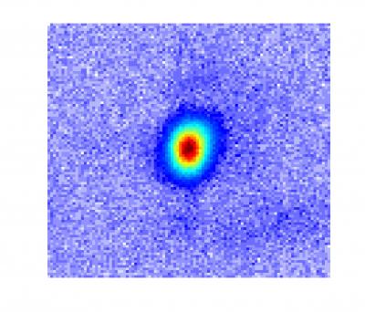 |
| This picture shows an actual image of a coherent (laser-like) X-ray beam. In contrast to the incoherent (light-bulb-like) light emitted in all directions from a Roentgen X-ray tube, the X-rays produced by high harmonic generation emerge as well-directed, laser-like, beams. |
"Just as a violin or guitar string will emit harmonics of its fundamental sound tone when plucked strongly, an atom can also emit harmonics of light when plucked violently by a laser pulse," adds Murnane. "The laser pulse first plucks electrons from the atoms, before driving them back again where they can collide with the atoms from which they came. Any excess energy is emitted as high-energy ultraviolet photons."
Like many phenomena, when HHG was first discovered, there was little science to explain it, and it was considered more a curious phenomenon than a potentially useful light source. After years of work, scientists eventually understood how very high harmonics were emitted, however there was one major challenge that most researchers gave up on--for most wavelengths in the X-ray region, the output HHG beams were extremely weak.
Murnane, Kapteyn and their students realized that there might be a chance to overcome that challenge and turn HHG into a useful X-ray light source--the tabletop-scale X-ray laser that has been a goal for laser science since shortly after the laser was first demonstrated in 1960.
"This was not an easy task," says Murnane. "Unlike a laser--which gets more intense as more energy is pumped into the system--in HHG, if the laser hits the atoms too hard, too many electrons are liberated from the gas atoms, and those electrons cause the laser light to speed up. If the speed of the laser and X-rays do not match, there is no way to combine the many X-ray waves together to create a bright output beam, since the X-ray waves from different gas atoms will interfere destructively."
Popmintchev and JILA graduate student Ming-Chang Chen worked out conditions that enable X-ray waves from many atoms in the gas to interfere constructively. The key was to use a relatively long-wavelength, mid-infrared laser and a high pressure gas cell that also guides the laser light. The resulting bright, X-ray beams maintain the coherent, directed beam qualities of the laser that drives the process.
The HHG process is effective only when the atoms are hit "hard and fast" by the laser pulses, with durations nearing 10-14 seconds--a fundamental limit representing just a few oscillations of the electromagnetic fields. Murnane and Kapteyn pioneered the technology for generating such light pulses in the 1990s, and used those lasers to develop and utilize HHG-based light sources in the extreme-ultraviolet (EUV) region of the spectrum in the 2000s. However, while researchers were using those lasers and the HHG technique to measure ever-shorter duration light pulses, they were stymied in how to make coherent light at shorter wavelengths in the more penetrating X-ray region of the spectrum.
The new paper in Science, under lead author and senior research associate Popmintchev, demonstrates that breakthrough, showing that the understanding of the HHG process the researchers developed is broadly valid.
"We would have never found this if we hadn't sat down and thought about what happens overall during HHG, when we change the wavelength of the laser driving it, what parameters have to be changed to make it work," added Kapteyn. "The amazing thing is that the physics seem to be panning out even over a very broad range of parameters. Usually in science you find a scaling rule that prevents you from making a dramatic jump, but in this case, we were able to generate 1.6 keV - each X-ray photon was generated from more than 5,000 infrared photons."
When the researchers first started to work with ultrafast, mid-infrared lasers just a few years ago, they actually made a step backwards and generated bright extreme-ultraviolet light of longer wavelengths than they used to achieve in the lab.
"However, we discovered a new regime that helped us to realize, just on paper, that we could make this giant step forward towards much shorter electromagnetic wavelengths and generate bright, laser-like, soft and hard X-rays," adds Popmintchev. "What the experiments were suggesting back then looked too good to be true! It seemed that Mother Nature has combined together, in the most simple and beautiful way, all the microscopic and macroscopic physics. Now, we are already at X-ray wavelengths as short as roughly 7.7 angstroms, and we do not know the limit."
To truly control the beam of photons, the researchers needed to understand the HHG process at the atomic level and how X-rays emitted from individual atoms combine to form a coherent beam of light.
That understanding combines microscopic and macroscopic models of the HHG process with the fact that those interactions occur at very high intensity in a dynamically changing medium. The development of such a conceptual understanding took the last decade to develop.
The result was the realization that there is no fundamental limit to the energy of the photons that can be generated using the HHG process. To obtain higher-energy photons, the system paradoxically begins with laser light using lower energy photons--specifically, mid-infrared lasers.
The JILA researchers demonstrated the validity of that principle in their labs in Colorado, but to achieve their breakthrough, the researchers traveled to Vienna with their beam-generating setup. There, they used a laser developed by co-author Andrius Baltuška and colleagues at the Vienna University of Technology--the world's most-intense ultrashort-pulse laser operating in the mid-infrared, with a wavelength of four microns.
"Thirty years ago, people were saying we could make a coherent X-ray source, but it would have to be an X-ray laser, and we'd need an atomic bomb as the energy source to pump it," said Deborah Jackson, the program officer who oversees the ERC's grant. "Now, we have these guys who understand the science fundamentals well enough to introduce new tricks for efficiently extracting energetic photons, pulling them out at X-ray wavelengths . . . and it's all done on a table-top!"
In addition to achieving the high energy, the increasingly broad spectrum opens a range of new applications.
"In an experiment using such a source, one energy region from the beam will correspond with one element, another with another element, and so on to simultaneously look at atoms across entire molecules, and that will allow us to see how charge moves from one part of a molecule to another as a chemical reaction is happening," adds Kapteyn. "It'll take us awhile to learn how to use this, but it's very exciting."
More information: "Bright Coherent Ultrahigh Harmonics in the keV X-ray Regime from Mid-Infrared Femtosecond Lasers," by T. Popmintchev et al, Science, 2012.
Wednesday, April 18, 2012
Looking through the glass transition on an ultrafast timescale
Glass is one of civilization's most valuable and versatile materials. To scientists, it's also one of the most intriguing, because it displays properties of both solids and liquids.
Glass is a non-crystalline solid that transforms into a liquid when it's heated to the so-called glass-transition temperature. When glass approaches this critical temperature—which is between 970 to 1,100 degrees Fahrenheit for the most common type of glass—it is simultaneously composed of fluid, flowing regions and solid-like, rigid domains.
For decades, scientists have been trying to understand exactly how glass behaves, at the molecular level, as it approaches the transition temperature. Now a research group led by University of Michigan chemist Kevin J. Kubarych has applied ultrafast spectroscopy to observe the fastest molecular motions of a liquid hovering just above its glass transition temperature.
"Progress in demystifying the glass transition can have a wide impact in many other fields, including predicting optical and mechanical properties of polymers and understanding crowded cellular environments of living organisms," said Kubarych, an assistant professor of chemistry.
Working with U-M chemistry graduate students John King and Matthew Ross, Kubarych found that even on the time scale of picoseconds there are signatures of "dynamic arrest": The molecules become locked into their positions and long-range motion grinds to a near halt, though, structurally, the glass is indistinguishable from a liquid.
Typically, these effects are observed on much slower time scales of seconds, minutes, or even longer. A paper summarizing the research was published online April 9 in Physical Review Letters. King is the first author of the paper.
More information: Ultrafast α-Like Relaxation of a Fragile Glass-Forming Liquid Measured Using Two-Dimensional Infrared Spectroscopy
Sunday, April 01, 2012
New technique lights up the creation of holograms
 The mechanism of full-color holography The major difference between the researchers’ technique and ordinary holography is the use of a prism to adjust the light beam’s angle of incidence in combination with a thin silver film to produce the surface plasmons. When the angle of incidence is controlled appropriately, the silver film is excited to produce surface plasmons, which in turn cause the white light to reach the hologram in the three primary colors of red, green and blue, producing a floating full-color stereoscopic image. Although only still images can be obtained at present, Kawata is planning to improve the current system to enable movie imaging based on the same principle in the future.
Thursday, March 29, 2012
Ultrafast laser pulses shed light on elusive superconducting mechanism
An international team that includes University of British Columbia physicists has used ultra-fast laser pulses to identify the microscopic interactions that drive high-temperature superconductivity.
In the experiment, to be outlined this Friday in the journal Science, electrons in a prototypical copper-oxide superconductor were excited by extremely short 100-femtosecond laser pulses.
As the material's electrons relax back to an equilibrium state, they release their excess energy via deformation of the superconductor's atomic lattice (phonons) or perturbation of its magnetic correlations (spin fluctuations).
The researchers were able to capture very fine grained data on the speed of the relaxation process and its influence on the properties of the superconducting system, showing that the high-critical temperature of these compounds can be accounted for by purely electronic (magnetic) processes.
"This new technique offers us our best window yet on the interactions that govern the formation of these elusive superconducting properties--both across time and across a wide range of characteristic energies," says UBC Associate Professor Andrea Damascelli, Canada Research Chair in Electronic Structure of Solids with the Department of Physics and Astronomy and the UBC Quantum Matter Institute.
"We're now able to begin to disentangle the different interactions that contribute to this fascinating behavior."
Superconductivity--the phenomenon of conducting electricity with no resistance--occurs in some materials at very low temperatures. High-temperature cuprate superconductors are capable of conducting electricity without resistance at temperatures as high as -140 degrees Celsius.
The key mechanism that allows the carriers to flow without resistance in superconductors stems from an effective pairing between electrons. In conventional metallic superconductors, this pairing mechanism is well understood as phonon-mediated. In copper-oxides, the nature of the low-resistance interaction between the electrons has remained a mystery.
"This breakthrough in the understanding of the puzzling properties of copper-oxides paves the way to finally solving the mystery of high-temperature superconductivity and revealing the key knobs for engineering new superconducting materials with even higher transition temperatures," says the paper's lead author Claudio Giannetti, a researcher with Italy's Università Cattolica del Sacro Cuore and visiting professor at UBC's Quantum Matter Institute.
The international collaboration also involved contributions from Japanese, Swiss and American researchers.
More information: "Disentangling the Electronic and Phononic Glue in a High-Tc Superconductor," by S. Dal Conte, et al., Science.
In the experiment, to be outlined this Friday in the journal Science, electrons in a prototypical copper-oxide superconductor were excited by extremely short 100-femtosecond laser pulses.
As the material's electrons relax back to an equilibrium state, they release their excess energy via deformation of the superconductor's atomic lattice (phonons) or perturbation of its magnetic correlations (spin fluctuations).
The researchers were able to capture very fine grained data on the speed of the relaxation process and its influence on the properties of the superconducting system, showing that the high-critical temperature of these compounds can be accounted for by purely electronic (magnetic) processes.
"This new technique offers us our best window yet on the interactions that govern the formation of these elusive superconducting properties--both across time and across a wide range of characteristic energies," says UBC Associate Professor Andrea Damascelli, Canada Research Chair in Electronic Structure of Solids with the Department of Physics and Astronomy and the UBC Quantum Matter Institute.
"We're now able to begin to disentangle the different interactions that contribute to this fascinating behavior."
Superconductivity--the phenomenon of conducting electricity with no resistance--occurs in some materials at very low temperatures. High-temperature cuprate superconductors are capable of conducting electricity without resistance at temperatures as high as -140 degrees Celsius.
The key mechanism that allows the carriers to flow without resistance in superconductors stems from an effective pairing between electrons. In conventional metallic superconductors, this pairing mechanism is well understood as phonon-mediated. In copper-oxides, the nature of the low-resistance interaction between the electrons has remained a mystery.
"This breakthrough in the understanding of the puzzling properties of copper-oxides paves the way to finally solving the mystery of high-temperature superconductivity and revealing the key knobs for engineering new superconducting materials with even higher transition temperatures," says the paper's lead author Claudio Giannetti, a researcher with Italy's Università Cattolica del Sacro Cuore and visiting professor at UBC's Quantum Matter Institute.
The international collaboration also involved contributions from Japanese, Swiss and American researchers.
More information: "Disentangling the Electronic and Phononic Glue in a High-Tc Superconductor," by S. Dal Conte, et al., Science.
Thursday, March 22, 2012
NIF facility fires record laser shot into target chamber
(PhysOrg.com) -- The National Ignition Facility (NIF) at Lawrence Livermore National Laboratory in California has set a new record for a laser shot. This past week, its combined 192 lasers fired a single 1.875-megajoule shot into an empty test chamber. After passing through the last of its focusing lens, the shot reached 2.03 megajoules, making it the first 2 megajoule ultraviolet laser.
Prior to this achievement, the most the facility had managed to coax out of the laser, the world’s largest, was 1.6 megajoules. Also, the new record shows that the NIF laser is capable of producing more than it was designed for, which was 1.8 megajoules. It also proved that it was capable of doing so without damaging its parts, allowing for another shot a day and a half later, which is important, because one of the goals for the laser is to get it to fire off shots at 15 per second eventually. That’s what researchers think will be needed to produce power economically from the laser system.
The ultimate goal of the NIF is to figure out a way to use a laser to produce nuclear fusion in a way that gets more energy out than is put in. Currently, that goal is still a ways off. Thus far, engineers at the project haven't even reached the break-even or ignition point, though they expect that to occur sometime this year. Tweaking the laser to produce more than it was designed for is a step in that direction. The NIF facility was designed to produce a fusion reaction by imploding hydrogen isotope pellets using the huge laser. To that end, the team has made steady progress. When the project first began eighteen months ago, it had just one percent of conditions in place that are believed necessary to achieve the ignition point. They have improved that mark to ten percent and it’s because the pace has picked up dramatically in recent months that they believe they will achieve the ignition point sometime over the next six months, which is when the original ignition campaign was slated to end.
Because the facility is funded by the US nuclear weapons complex, there has been debate about whether it would ever be used to prove or disprove the idea that lasers could be used to create nuclear fusion to produce electric power. Having the laser break records doesn’t really resolve that argument in the short term, but it might in the long run if it does eventually show that electricity could be created economically using such a process.
More information: The National Ignition Facility (NIF): https://lasers.llnl.gov/
Prior to this achievement, the most the facility had managed to coax out of the laser, the world’s largest, was 1.6 megajoules. Also, the new record shows that the NIF laser is capable of producing more than it was designed for, which was 1.8 megajoules. It also proved that it was capable of doing so without damaging its parts, allowing for another shot a day and a half later, which is important, because one of the goals for the laser is to get it to fire off shots at 15 per second eventually. That’s what researchers think will be needed to produce power economically from the laser system.
The ultimate goal of the NIF is to figure out a way to use a laser to produce nuclear fusion in a way that gets more energy out than is put in. Currently, that goal is still a ways off. Thus far, engineers at the project haven't even reached the break-even or ignition point, though they expect that to occur sometime this year. Tweaking the laser to produce more than it was designed for is a step in that direction. The NIF facility was designed to produce a fusion reaction by imploding hydrogen isotope pellets using the huge laser. To that end, the team has made steady progress. When the project first began eighteen months ago, it had just one percent of conditions in place that are believed necessary to achieve the ignition point. They have improved that mark to ten percent and it’s because the pace has picked up dramatically in recent months that they believe they will achieve the ignition point sometime over the next six months, which is when the original ignition campaign was slated to end.
Because the facility is funded by the US nuclear weapons complex, there has been debate about whether it would ever be used to prove or disprove the idea that lasers could be used to create nuclear fusion to produce electric power. Having the laser break records doesn’t really resolve that argument in the short term, but it might in the long run if it does eventually show that electricity could be created economically using such a process.
More information: The National Ignition Facility (NIF): https://lasers.llnl.gov/
Thursday, March 15, 2012
Scientists reveal inner workings of magnets, a finding that could lead to faster computers
Using a light source that creates X-ray pulses only one quadrillionth of a second in duration, the Boulder team was able to observe how magnetism in nickel and iron atoms works, and they found that each metal behaves differently. One quadrillionth of a second is a million times faster than one billionth of a second.
The results of the study were published online this week by the Proceedings of the National Academy of Sciences. Six of the study’s 19 co-authors are located at CU-Boulder.
Many technology experts believe that next-generation computer disk drives will use optically-assisted magnetic recording to achieve much higher drive capacities, according to NIST scientist Tom Silva, who worked with CU-Boulder physics professors Margaret Murnane and Henry Kapteyn on the research. However, many questions remain about how the delivery of optical energy to the magnetic system can be optimized for maximum drive performance. And this finding could help researchers answer some of their questions.
“The discovery that iron and nickel are fundamentally different in their interaction with light at ultrafast time scales suggests that the magnetic alloys in hard drives could be engineered to enhance the delivery of the optical energy to the spin system,” Silva said.
Magnetism exists because all of the “spins” in a magnet -- each of which is like a very small bar magnet with a north and south pole -- are lined up to point in the same direction, much like members of a marching band who are moving in unison, explained Murnane, who also is a fellow of JILA, a joint institute of CU-Boulder and NIST.
“The powerful laser pulse scrambles the magnetic spins in the metal, as if the members of the marching band started moving in different directions across the football field, causing the magnetization to rapidly disappear within a mere fifty quadrillionths of a second, a process known as ultrafast demagnetization,” Murnane said.
While ultrafast demagnetization has been a well-known phenomenon since its discovery in 1996, the CU and NIST researchers saw for the first time that different kinds of spins in metal scramble on different time scales. Until now, it was assumed that all the spins in a metal alloy behaved in the same way due to a powerful quantum mechanical effect known as the exchange interaction, which lines up all the individual spins in the same direction.
“What we have seen for the first time is that the iron spins and the nickel spins react to light in different ways, with the iron spins being mixed up by light much more readily than the nickel spins,” said Silva. “In the end, the exchange interaction still pulls the two spin systems back into synchronization after a few quadrillionths of a second. Seeing such a difference was only possible by taking advantage of the extremely fast X-ray technology developed at the University of Colorado and elsewhere.”
The laser technology used in the experiment, known as “high harmonic generation,” can generate laser-like beams of X-rays that span a wide portion of the electromagnetic spectrum, including the spectral region where nickel and iron interact very strongly with X-rays.
The results of the study were published online this week by the Proceedings of the National Academy of Sciences. Six of the study’s 19 co-authors are located at CU-Boulder.
Many technology experts believe that next-generation computer disk drives will use optically-assisted magnetic recording to achieve much higher drive capacities, according to NIST scientist Tom Silva, who worked with CU-Boulder physics professors Margaret Murnane and Henry Kapteyn on the research. However, many questions remain about how the delivery of optical energy to the magnetic system can be optimized for maximum drive performance. And this finding could help researchers answer some of their questions.
“The discovery that iron and nickel are fundamentally different in their interaction with light at ultrafast time scales suggests that the magnetic alloys in hard drives could be engineered to enhance the delivery of the optical energy to the spin system,” Silva said.
Magnetism exists because all of the “spins” in a magnet -- each of which is like a very small bar magnet with a north and south pole -- are lined up to point in the same direction, much like members of a marching band who are moving in unison, explained Murnane, who also is a fellow of JILA, a joint institute of CU-Boulder and NIST.
“The powerful laser pulse scrambles the magnetic spins in the metal, as if the members of the marching band started moving in different directions across the football field, causing the magnetization to rapidly disappear within a mere fifty quadrillionths of a second, a process known as ultrafast demagnetization,” Murnane said.
While ultrafast demagnetization has been a well-known phenomenon since its discovery in 1996, the CU and NIST researchers saw for the first time that different kinds of spins in metal scramble on different time scales. Until now, it was assumed that all the spins in a metal alloy behaved in the same way due to a powerful quantum mechanical effect known as the exchange interaction, which lines up all the individual spins in the same direction.
“What we have seen for the first time is that the iron spins and the nickel spins react to light in different ways, with the iron spins being mixed up by light much more readily than the nickel spins,” said Silva. “In the end, the exchange interaction still pulls the two spin systems back into synchronization after a few quadrillionths of a second. Seeing such a difference was only possible by taking advantage of the extremely fast X-ray technology developed at the University of Colorado and elsewhere.”
The laser technology used in the experiment, known as “high harmonic generation,” can generate laser-like beams of X-rays that span a wide portion of the electromagnetic spectrum, including the spectral region where nickel and iron interact very strongly with X-rays.
Saturday, March 10, 2012
Making sharper X-rays
 A photograph of the custom-built laser chamber, which receives simultaneously a pulse of helium gas and a pulse from RIKEN’s free-electron laser, which is known as the SPring-8 Compact SASE Source (SCSS). The resulting blue-green superfluorescence is visible.
A variety of imaging technologies rely on light with short wavelengths because it allows very small structures to be resolved. However, light sources which produce short, extreme ultraviolet or x-ray wavelengths often have unstable emission wavelength and timing. Now, by illuminating a gas with a powerful laser, a research team in Japan has demonstrated a light source that may solve many of these problems. The research was published by Mitsuru Nagasono, from the RIKEN SPring-8 Center in Harima, and his colleagues from RIKEN and three other institutes.
More information: Nagasono, M., et al. Observation of free-electron-laser-induced collective spontaneous emission (superfluorescence). Physical Review Letters 107, 193603 (2011).
Friday, March 09, 2012
Femtolaser Pulse Creates 3-D Nanostructures
A new fabrication process using femtosecond lasers creates 3-D nanostructures in materials, an essential step toward creating invisibility cloaks and other advanced materials that bend light in unusual ways.
Researchers in Eric Mazur's laboratory at the Harvard School of Engineering and Science (SEAS) fired a femtosecond laser, which releases incredibly bright flashes of light that last 5 x 10^-14, s, at a glass slide coated with a mixture of silver nitrate, water and PVP, a water-soluble polymer. The laser blast changes the electrical, physical and optical properties of the slide, and photoreduces the silver ions on the slide into nanocrystals of silver metal suspended on the polymer.
Previous attempts to create a 3-D structure failed because the coating was not quite right. When the researchers used only the silver nitrate and water, there was no lattice support for the silver atoms, they said.
"Normally, when people use femtosecond lasers in fabrication, they’re creating a woodpile structure: something stacked on something else, being supported by something else. If you want to make an array of silver dots, however, they can’t float in space," Mazur said. Ethanol and the PVP polymer were added to the solution to provide support to the structure, but reactions were fast and uncontrollable. Removing the ethanol solved the problem entirely.
"What was most surprising about it was how simple it is. It was a matter of using less," Mazur said.
The new fabrication process advances nanoscale metal lithography into three dimensions, and does it at a resolution high enough to be practical for metamaterials.
"This work demonstrates that we can create silver dots that are disconnected in X, Y, and Z," said Kevin Vora, a graduate student working on the project. "There’s no other technique that feasibly allows you to do that."
The work, which was supported by the Air Force Office of Scientific Research, is described in Applied Physics Letters.
Researchers in Eric Mazur's laboratory at the Harvard School of Engineering and Science (SEAS) fired a femtosecond laser, which releases incredibly bright flashes of light that last 5 x 10^-14, s, at a glass slide coated with a mixture of silver nitrate, water and PVP, a water-soluble polymer. The laser blast changes the electrical, physical and optical properties of the slide, and photoreduces the silver ions on the slide into nanocrystals of silver metal suspended on the polymer.
Previous attempts to create a 3-D structure failed because the coating was not quite right. When the researchers used only the silver nitrate and water, there was no lattice support for the silver atoms, they said.
"Normally, when people use femtosecond lasers in fabrication, they’re creating a woodpile structure: something stacked on something else, being supported by something else. If you want to make an array of silver dots, however, they can’t float in space," Mazur said. Ethanol and the PVP polymer were added to the solution to provide support to the structure, but reactions were fast and uncontrollable. Removing the ethanol solved the problem entirely.
"What was most surprising about it was how simple it is. It was a matter of using less," Mazur said.
The new fabrication process advances nanoscale metal lithography into three dimensions, and does it at a resolution high enough to be practical for metamaterials.
"This work demonstrates that we can create silver dots that are disconnected in X, Y, and Z," said Kevin Vora, a graduate student working on the project. "There’s no other technique that feasibly allows you to do that."
The work, which was supported by the Air Force Office of Scientific Research, is described in Applied Physics Letters.
Thursday, March 08, 2012
First-ever images of atoms moving in a molecule captured
 Researchers at Ohio State University and Kansas State University have captured the first-ever images of atoms moving in a molecule. Shown here is molecular nitrogen. The researchers used an ultrafast laser to knock one electron from the molecule, and recorded the diffraction pattern that was created when the electron scattered off the molecule. The image highlights any changes the molecule went through during the time between laser pulses: one quadrillionth of a second. The constituent atoms' movement is shown as a measure of increasing angular momentum, on a scale from dark blue to pink, with pink showing the region of greatest momentum. Credit: Image courtesy of Cosmin Blaga, Ohio State University.
Key to the experiment, which appears in this week's issue of the journal Nature, is the researchers' use of the energy of a molecule's own electron as a kind of "flash bulb" to illuminate the molecular motion.
The team used ultrafast laser pulses to knock one electron out of its natural orbit in a molecule. The electron then fell back toward the molecule scattered off of it, analogous to the way a flash of light scatters around an object, or a water ripple scatters in a pond.
Principal investigator Louis DiMauro of Ohio State University said that the feat marks a first step toward not only observing chemical reactions, but also controlling them on an atomic scale.
"Through these experiments, we realized that we can control the quantum trajectory of the electron when it comes back to the molecule, by adjusting the laser that launches it," said DiMauro, who is a professor of physics at Ohio State. "The next step will be to see if we can steer the electron in just the right way to actually control a chemical reaction."
A standard technique for imaging a still object involves shooting the object with an electron beam – bombarding it with millions of electrons per second. The researchers' new single-electron quantum approach allowed them to image rapid molecular motion, based on theoretical developments by the paper's coauthors at Kansas State University.
A technique called laser induced electron diffraction (LIED) is commonly used in surface science to study solid materials. Here, the researchers used it to study the movement of atoms in a single molecule.
The molecules they chose to study were simple ones: nitrogen, or N2, and oxygen, or O2. N2 and O2 are common atmospheric gases, and scientists already know every detail of their structure, so these two very basic molecules made a good test case for the LIED method.
In each case, the researchers hit the molecule with laser light pulses of 50 femtoseconds, or quadrillionths of a second. They were able to knock a single electron out of the outer shell of the molecule and detect the scattered signal of the electron as it re-collided with the molecule.
DiMauro and Ohio State postdoctoral researcher Cosmin Blaga likened the scattered electron signal to the diffraction pattern that light forms when it passes through slits. Given only the diffraction pattern, scientists can reconstruct the size and shape of the slits. In this case, given the diffraction pattern of the electron, the physicists reconstructed the size and shape of the molecule – that is, the locations of the constituent atoms' nuclei.
The key, explained Blaga, is that during the brief span of time between when the electron is knocked out of the molecule and when it re-collides, the atoms in the molecules have moved. The LIED method can capture this movement, "similar to making a movie of the quantum world," he added.
Beyond its potential for controlling chemical reactions, the technique offers a new tool to study the structure and dynamics of matter, he said. "Ultimately, we want to really understand how chemical reactions take place. So, long-term, there would be applications in materials science and even chemical manufacturing."
"You could use this to study individual atoms," DiMauro added, "but the greater impact to science will come when we can study reactions between more complex molecules. Looking at two atoms – that's a long way from studying a more interesting molecule like a protein."
Subscribe to:
Posts (Atom)